

Pulmonary nodules identified in patient with neurological symptoms
Patient data
A male patient known to be hypertensive, with peripheral vascular disease, is brought to the hospital by emergency for expressive language disorders that started 2 days ago. In evolution, the patient's clinical condition worsens progressively with the appearance of respiratory failure and the impairment of the state of consciousness.
Case discussion
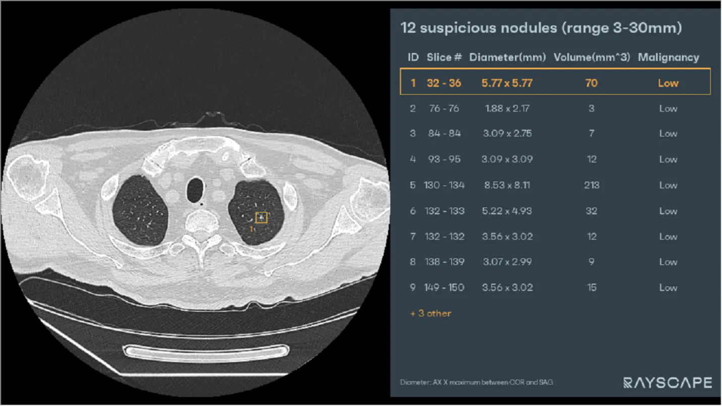
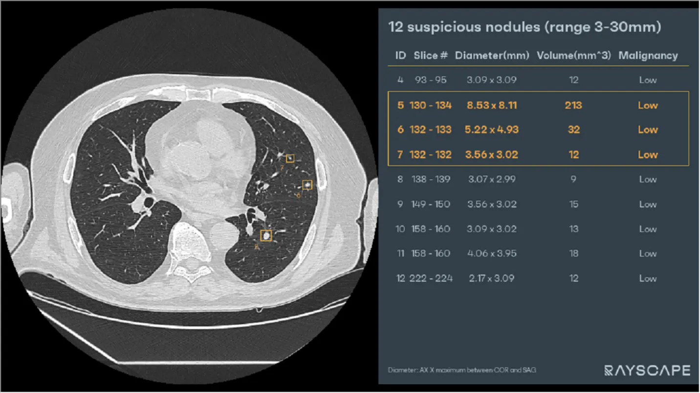
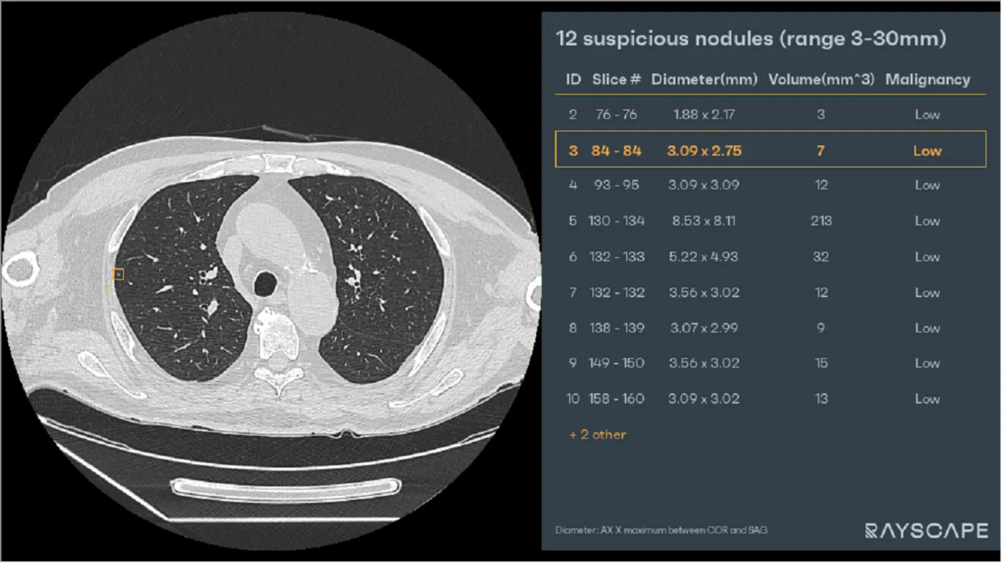
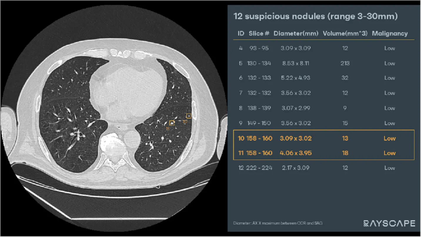
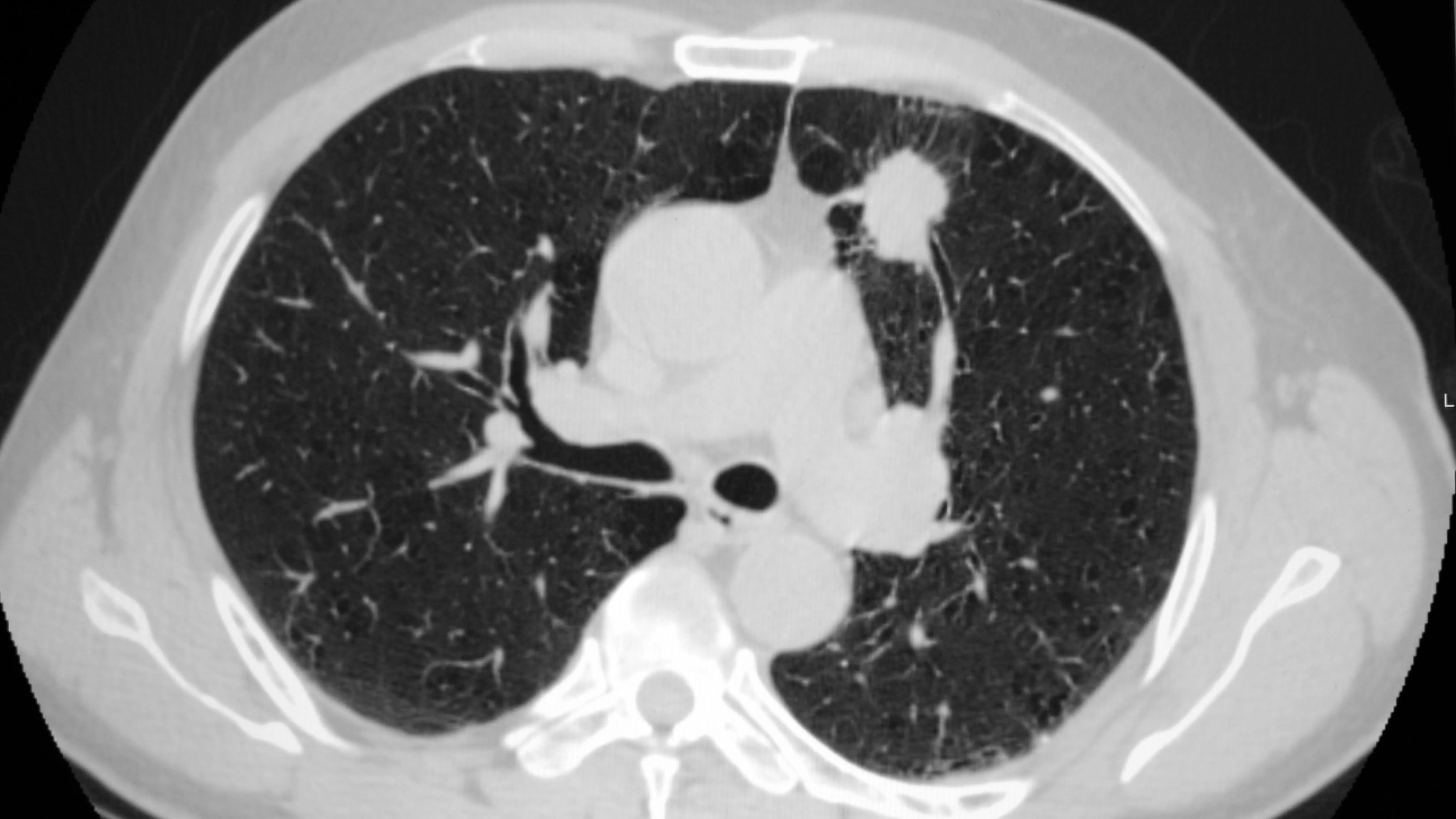
Due to the unstable condition at presentation, following the explorations performed for the diagnosis and treatment of the primary neurological condition, a series of pulmonary imaging explorations in the form of lung CTs are also carried out. The Rayscape algorithms detect multiple pulmonary nodules which are then confirmed by the radiologist. The biggest nodules have diameters of 6 mm and 8 mm, respectively, and are located at the level of the upper left lobe and the lower left lobe.
If initially the attention was focused on the neurological symptoms, after the treatment, the findings at the pulmonary level gained greater importance for the patient's future state of health.
This is an example of a case where the pathology of a patient is signaled by the algorithm during the exploration for the etiology of other possible diseases, which could go easily unnoticed. The patient was referred for a multidisciplinary surgical and oncological consultation.
More info:
Finding out if a lung nodule is malignant or benign is the main goal of the diagnostic procedure. The nodule’s development rate can be used as a reliable indicator of the difference. Benign nodules rarely grow in size. In comparison, malignant nodules might grow by a factor of two every four months on average (some as quickly as 25 days, some as slowly as 15 months). A sequence of X-rays or CT (computed tomography) images taken over a period can be used to assess the growth rate.
The second most reliable approach to tell if it is malignant or benign is evaluating a nodule’s calcification. Nodules that are benign have a tendency to be more uniformly colored, smoother, and more consistently formed. Nodules that are cancerous are much more prone to have asymmetrical forms, rougher surfaces, and varying colors or spotted patterns like granular, punctate or reticular patterns of calcification.
An imaging finding of calcification in a lung nodule suggests that the lesion is likely benign. However, not all calcified pulmonary nodules are benign, and main central lung carcinoid, metastasis, and primary bronchogenic cancer are among the differential diagnoses. The ability to identify calcification in malignant tumors is now more sensitive because to the widespread use of computed tomography.
The primary radiographic characteristic is that lung nodules are often homogenous and well-defined, because of their sharp borders with normal aerated lung parenchyma surrounding them. Lung nodules may be solitary or multiple. To be considered solitary, a nodule must be completely surrounded by normal lung parenchyma, without associated atelectasis, enlargement of the hilum, or pleural effusion.
X-rays or CT scans often give enough details to make an accurate diagnosis. The nodule’s cells might be collected by medical professionals for a biopsy. Using a needle or by conducting local surgery, cells can be isolated and, in addition with a patient’s sputum analysis, it might offer more reliable information.
References:
1. Knipe, Henry. "Coin lesion (lung) | Radiology Reference Article | Radiopaedia.org". Radiopaedia.
2. Margerie-Mellon, Constance; Bankier, Alexander A. (1 December 2019). "To Be or Not to Be … a Pulmonary Nodule". Radiology: Cardiothoracic Imaging. 1 (5): e190201. doi:10.1148/ryct.2019190201. PMC 7977753. PMID 33778533.
3. Ost D, Fein AM, Feinsilver SH (June 2003). "Clinical practice. The solitary pulmonary nodule". The New England Journal of Medicine. 348 (25): 2535–2542. doi:10.1056/NEJMcp012290. PMID 12815140
4. https://xvision.app/blog/2022/07/27/evaluation-of-patients-with-pulmonary-nodules/

Discover how Rayscape works
integrate Rayscape AI into
your hospital's infrastructure
and improve both your
daily workflow and patients' experience.
