

Lung CT AI for Incidental Findings
If they’re missed, it’s not because they were invisible. It’s because they were outside the mental map of the case.
Not all lung findings start with a lung-focused scan. Some reveal themselves incidentally—on neck CTs, trauma protocols, or contrast studies ordered for other suspicions. And in these moments, subtle pulmonary nodules can be present, significant, and easily missed.
Next, you’ll follow incidental cases where Rayscape’s AI surfaced findings not being actively searched for—quiet markers that lived outside the scope of the original question, but not outside the patient’s story.
The reality of incidental findings
Modern practice includes:
- CTs for trauma, embolism, infection, surgical planning
- Partial field-of-view studies capturing only the upper or lower lungs
- Clinicians focused on non-pulmonary findings
- Radiologists facing time pressure and fragmented clinical context
And yet:
- Incidental nodules may fall outside the radiologist’s search pattern
- The Fleischner guidelines may not be applied consistently
- Subtle nodules risk being labeled “artifact” or “non-significant”
How AI makes a difference
- Reviews full lung volumes, even in partial-field scans
- Highlights nodules unrelated to original study indication
- Automatically applies Fleischner-based classification criteria
- Suggests structured language and consistent follow-up pathways
- Enhances awareness without interrupting primary reporting focus
Rayscape doesn’t change what’s being looked for—but it expands what can be seen. In the incidental, it restores intentionality.
Below, you’ll find real-world cases where incidental findings were surfaced thanks to Rayscape’s AI—cases where pulmonary nodules were not being actively searched for but could no longer be ignored.
Across these cases, Rayscape helped:
- Detect pulmonary nodules in CT scans performed for non-lung nodules indications
- Quantify and classify incidental lesions using Fleischner criteria
- Enable timely referral to follow-up
These are not hypothetical scenarios—they’re drawn from daily clinical flows, where attention is finite and the unexpected demands a second look.
#INCIDENTAL CASE 1: The signal behind the shock
Patient: Male, 51 years old
Setting: Emergency Room → Trauma CT
Clinical Presentation: High-speed collision, blunt chest trauma
Imaging: Contrast-enhanced trauma CT (head/chest/abdomen/pelvis)
Clinical Context
The scan wasn’t meant to find it.
The patient came in as a trauma code—classic blunt force scenario: tachycardia, shallow breathing, and a swelling hematoma across his right chest wall. The trauma bay moved on instinct. Portable X-ray confirmed a right pneumothorax, and the trauma CT followed before the ink dried on the consent.
And in that moment, the entire clinical mindset narrowed: Airway. Breathing. Circulation. Damage control.
The CT was loaded fast and read even faster—focused on rib fractures, hemothorax, any vascular breach. On the primary pass, that’s what the radiologist focused on too. Bilateral pleural effusions. A visible pneumothorax. No dissection, no tamponade, no abdominal bleed.
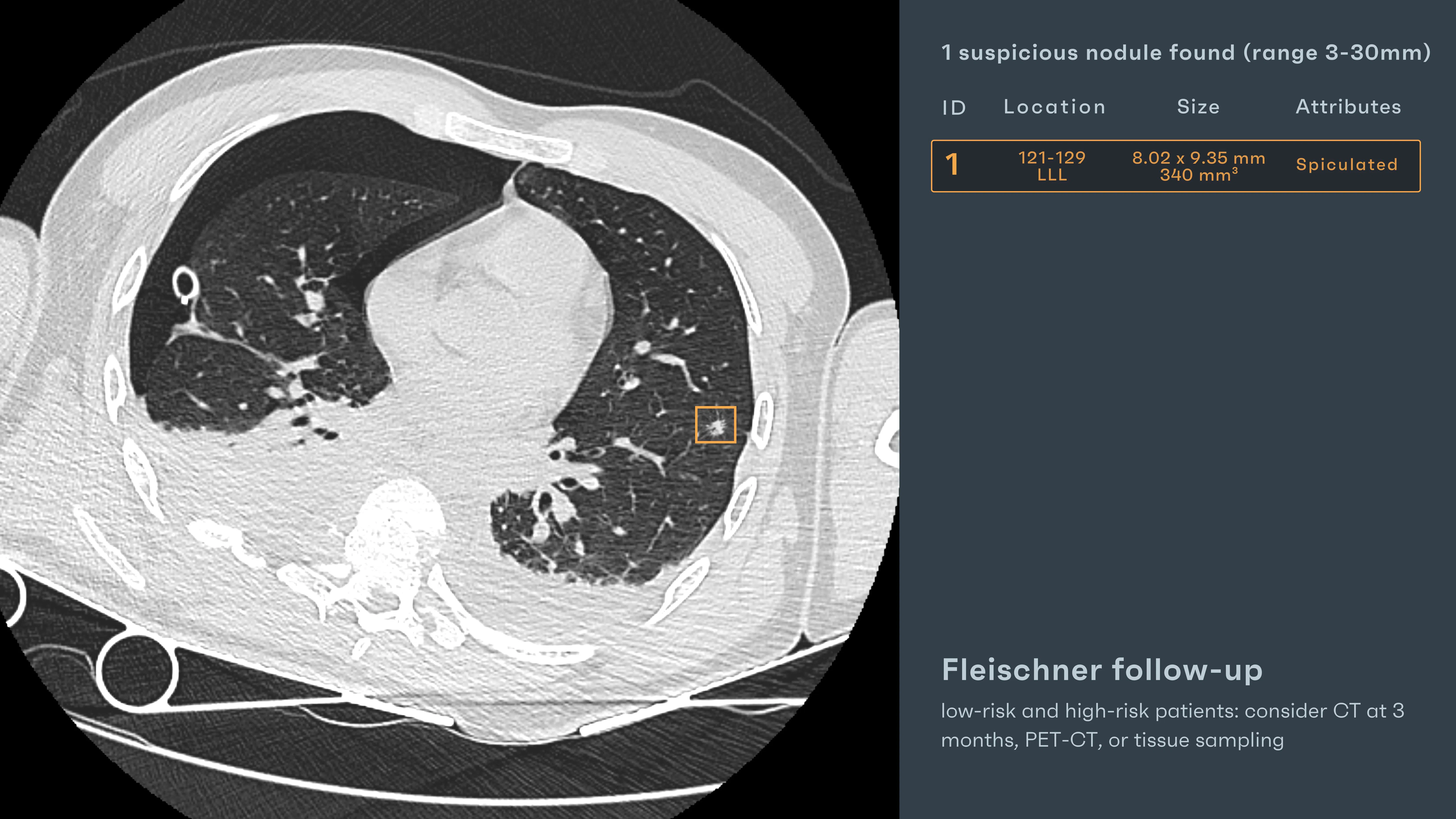
He could have cleared the scan with that. But something pulled at the corner of the screen—something not urgent, not dramatic, not life-threatening. Just a shadow in the left lower lobe. Angular. Not what they were looking for.
AI Interaction
While trauma injuries drew immediate clinical attention, Rayscape’s AI focused elsewhere—reviewing the lung parenchyma quietly in parallel.
- “Spiculated Nodule” – A solitary, dense lesion in the left lower lobe, measuring 8.02 x 9.35 mm, with a volume of 340 mm³.
- The AI detected lobulated margins, and spiculation—all consistent with potential malignancy.
Based on Fleischner Society guidelines, the system recommended: “CT at 3 months, PET-CT, or tissue sampling, considering both low- and high-risk patient profiles.”
This was not the finding of the day, but it might be the finding that matters most—the one no one asked about, and AI refused to ignore.
Outcome
The trauma was managed. The clinical documentation nearly closed. But the radiologist—alerted by Rayscape—paused at the left lower lobe. The nodule was added to the report with explicit follow-up. A re-evaluation CT was arranged.
That follow-up scan revealed growth. PET-CT and biopsy confirmed what AI had quietly warned: a malignant lesion, early-stage primary lung cancer. The trauma brought him in, but it was the incidental nodule—and the AI that surfaced it—that may have saved his life.
#INCIDENTAL CASE 2: What the margin contained
Patient: Female, 68 years old
Setting: Outpatient CT for mediastinal mass evaluation
Clinical Presentation: Referred for further characterization of a known anterior mediastinal lesion
Imaging: Contrast-enhanced Chest CT
Clinical Context
The request was focused: “Evaluate superior mediastinal” mass. That was the assignment—narrow, framed, clinically justified. And so, like many targeted scans, the eye moved first to the center: the mass. It was left-sided, well-marginated, and exactly where expected. Not invading, not urgent. This could’ve been a fast read.
One impression line. One follow-up recommendation. Done. But radiologists know: focus is a double-edged sword. What sharpens attention can also narrow it. What’s not in the question is often not in the answer.
AI Interaction
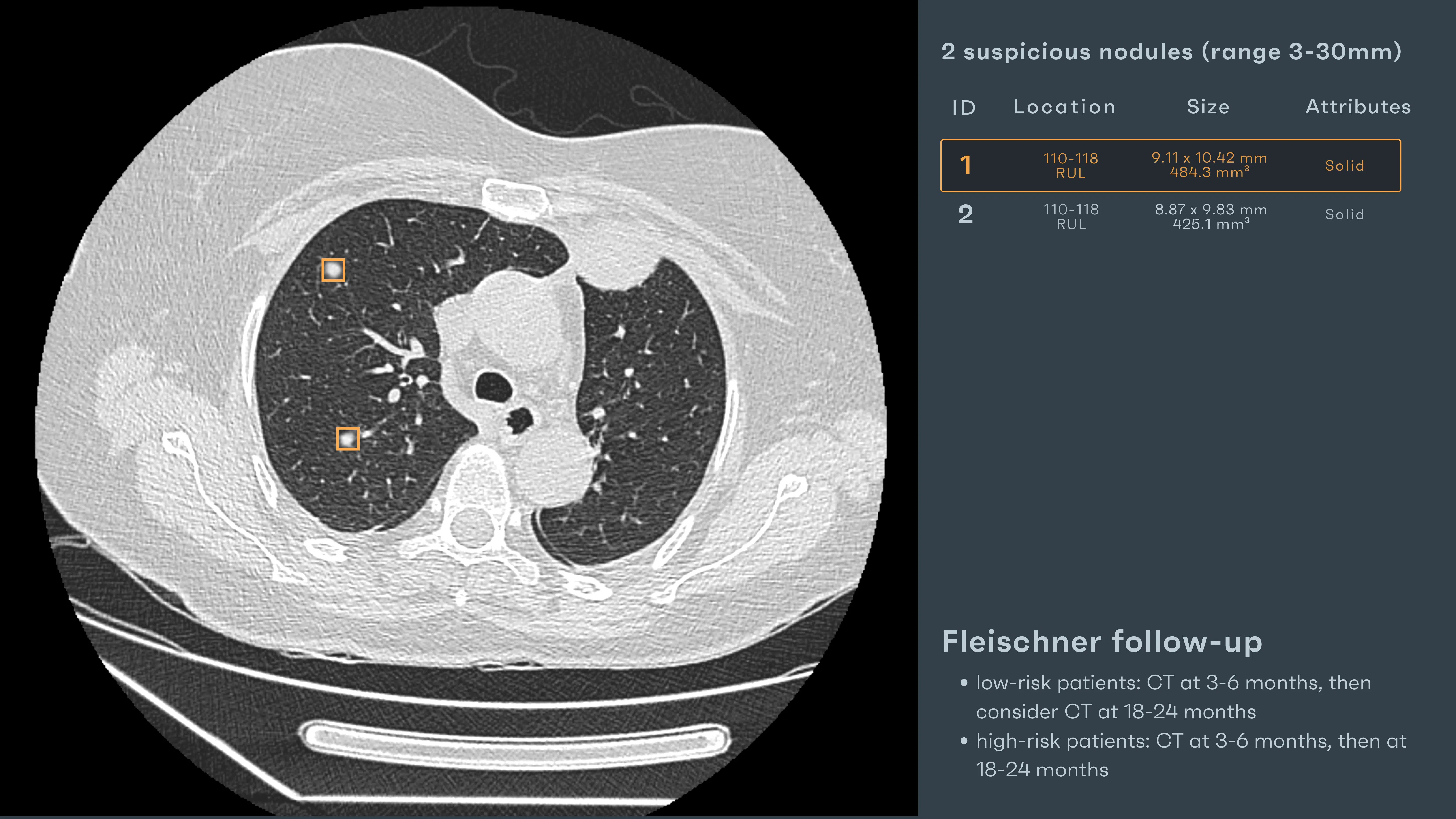
While the radiologist zoomed in on the mediastinal mass, Rayscape’s AI widened the scope—its gaze not limited by the referral note.
“Solid Nodule” – Right upper lobe:
- 9.11 x 10.42 mm, volume 484.3 mm³
- Good contour, discrete from the mediastinal pathology
“Second Solid Nodule” – Same lobe, nearby location:
- 8.87 x 9.83 mm, volume 425.1 mm³
- Slightly smoother margins, similar attenuation
Both findings were outside the immediate clinical target, but squarely within the AI’s review field. The Fleischner Society criteria were applied: “CT at 3–6 months, followed by 18–24 months for surveillance (regardless of risk category)”
Outcome
The mediastinal lesion was monitored as expected—stable over time, benign in behavior. But the nodules became the story. They were flagged in the final report, described, and scheduled for structured follow-up. At the 3-month scan, their size changed, no more stable in dimensions and volume. The AI didn’t find what was asked but it found what one could missed after a long day of work.
#INCIDENTAL CASE 3: The nodule outside the crisis
Patient: Male, 51 years old
Setting: Emergency Room – Acute Pulmonary Thromboembolism
Clinical Presentation: Sudden dyspnea, pleuritic chest pain, hypoxia
Imaging: Chest CT (contrast-enhanced)
Clinical Context
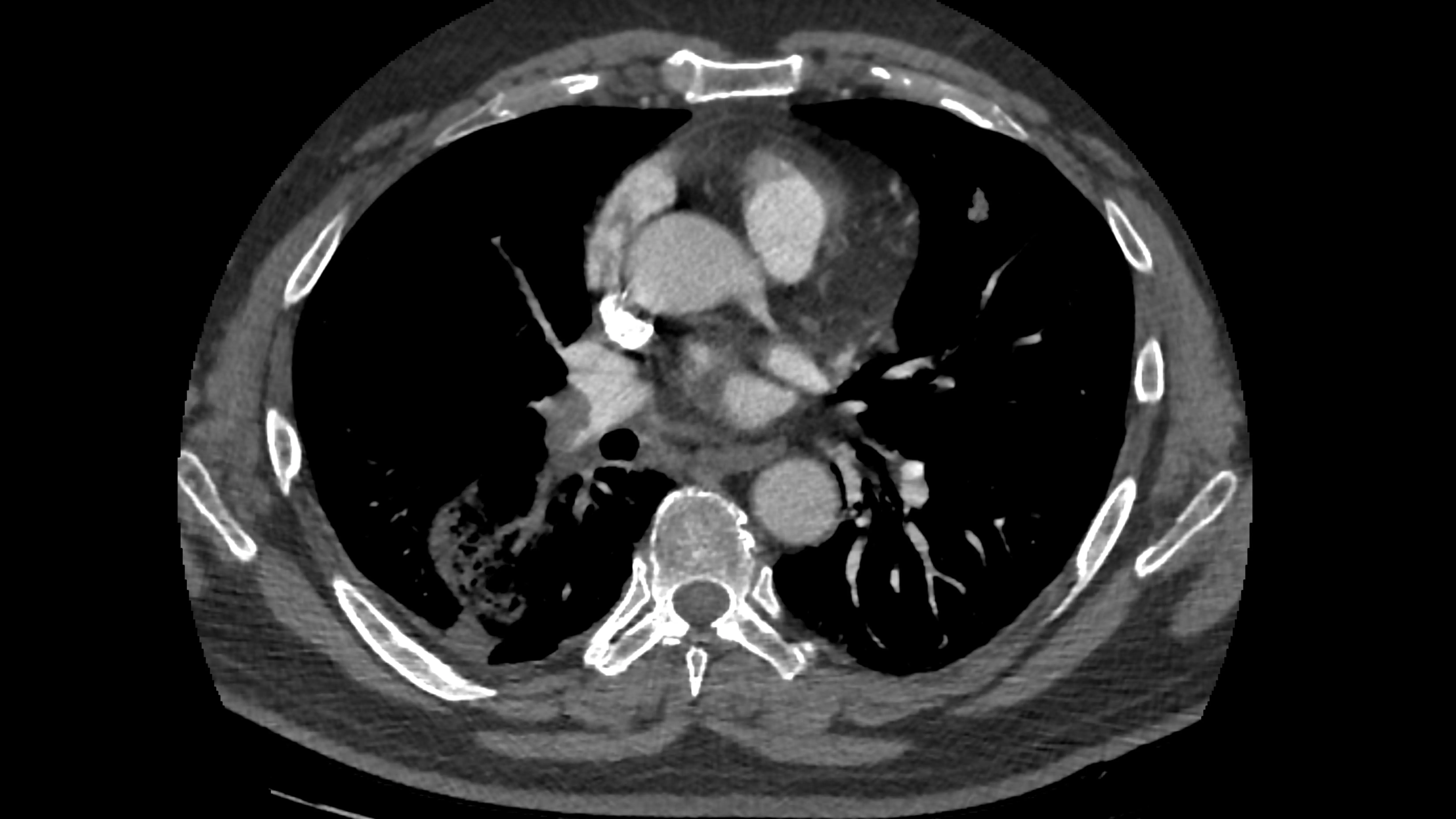
The diagnosis was urgent but clear: an acute pulmonary embolism. He had arrived at the ER gasping for air—saturation at 86%, tachycardic, diaphoretic. A stat CT angio was ordered, and it confirmed a clot in the right pulmonary artery, with wedge-shaped opacities in the right lower lobe suggesting infarction. The team mobilized: anticoagulation started, oxygen therapy adjusted.
In these high-pressure scenarios, the focus narrows—rightly—on the immediate threat.
AI Interaction
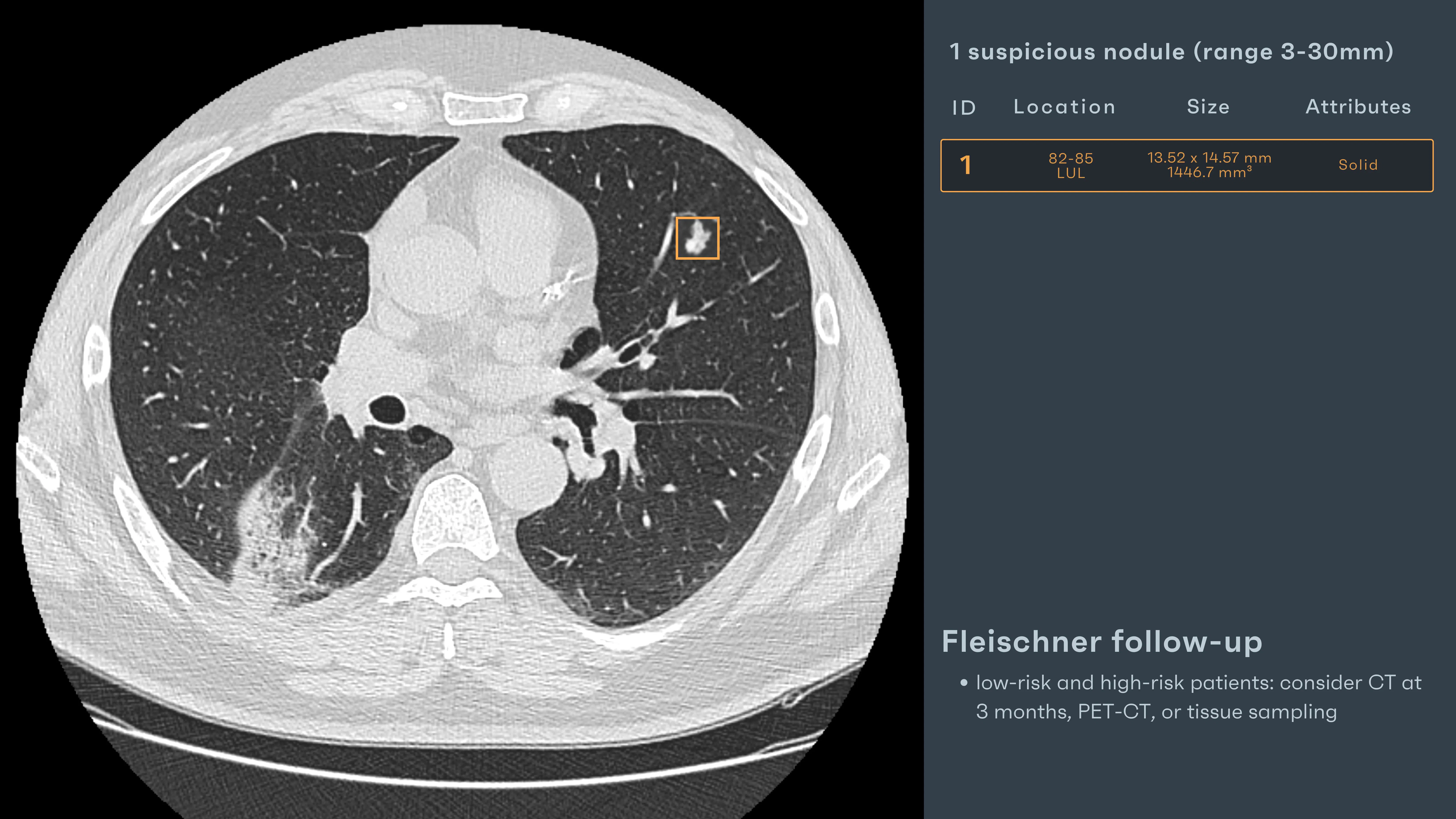
Rayscape AI processed the scan alongside the team. While the clot was being assessed and the infarct noted, the AI framed something else—in a different lobe, in a different tone:
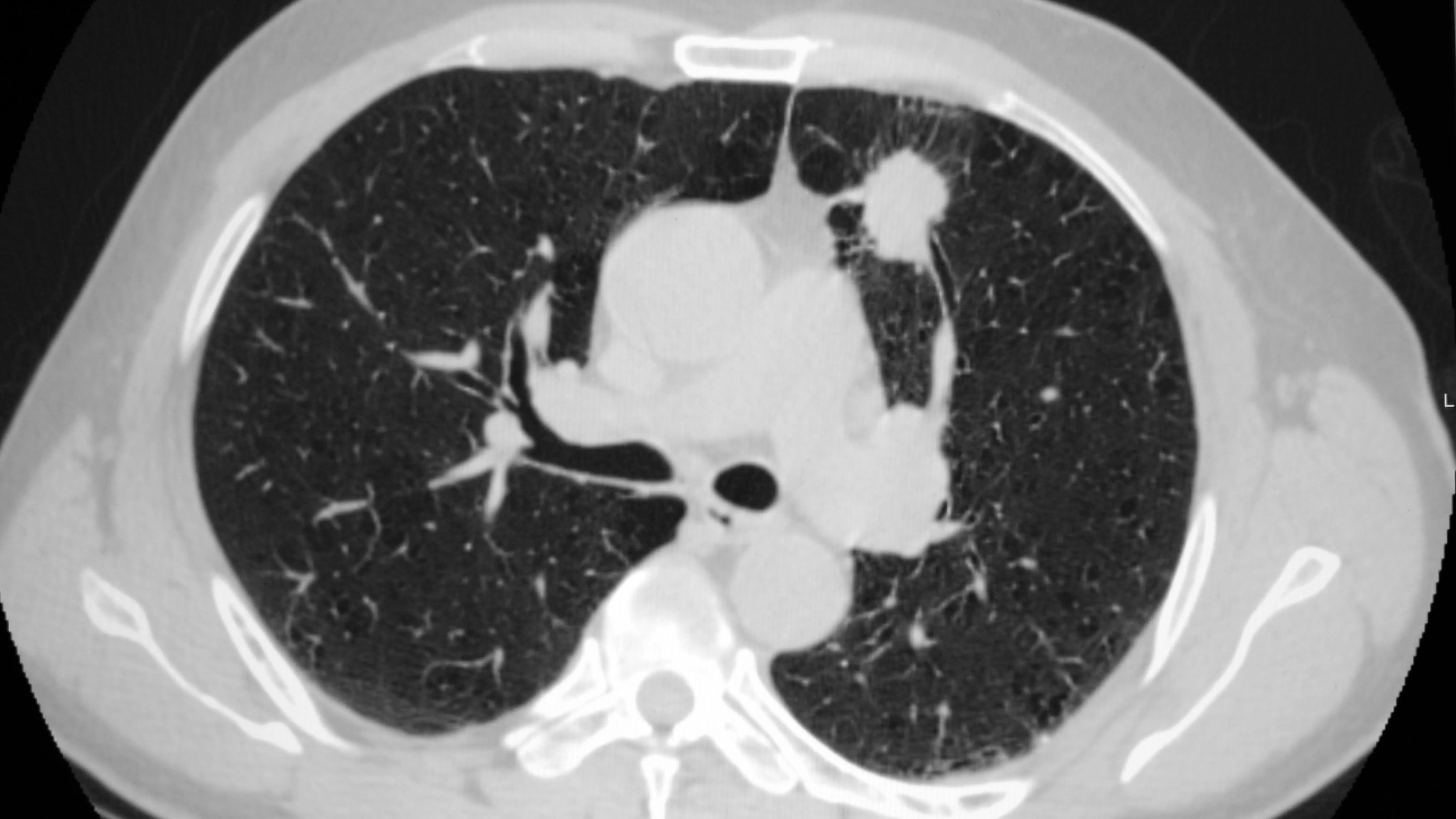
“Solid Nodule” – Left Upper Lobe (LUL)
- 13.52 x 14.57 mm, 1446.7 mm³
- Lobulated margins, distinct, not vascular
- Flagged for Fleischner follow-up: CT in 3 months, or consider PET-CT / biopsy
The finding had no relation to the PE. It was incidental—but not insignificant. And in an acute case, it may have gone entirely unnoticed without that overlay.
Outcome
The radiologist updated the report with a clear recommendation: “Incidental solid pulmonary nodule in LUL, lobulated, not consistent with infarction or inflammatory change. Recommend PET-CT and/or tissue sampling per Fleischner guidance.”
A PET-CT was arranged once the patient stabilized. The nodule showed intense uptake. Biopsy followed. Diagnosis: adenocarcinoma.
He had come in to treat a clot. Instead, AI surfaced a cancer—quiet, hidden, and potentially years from detection without this scan.
Discussions
Radiologists aren’t tireless or immune to overload. Behind every lung CT report is a reader juggling timelines, growth patterns, and nuanced findings—while the next scan waits in the queue. The pathology wasn’t hidden. It lived in ambiguity: a borderline nodule, a subtle ground-glass opacity, a shift just below the threshold of alarm.
The AI didn’t “catch” what the radiologist missed—it offered clarity before the fog could form. It positioned the findings within the frame, allowing the mind to move faster, the pattern to resolve more sharply. And this is the quiet revolution: not replacement, but reinforcement. Not autonomy, but augmentation. Rayscape acted not as a second opinion, but as a second set of steady hands in a moment where one pair may not be enough.
The future of lung CT is collaborative. Where human intuition meets algorithmic precision. Where a bounding box becomes not a conclusion, but a question, well-posed, in just the right place.
Built to fit your workflow
For IT teams and PACS administrators, implementation is often a key concern. Rayscape is designed to fit into your existing systems:
- Compatibility with major PACS platforms and cloud environments
- Vendor agnostic: trained to work on every DICOM image, no matter the vendor of the machine or the protocol
- Flexible deployment: on-premise, hybrid, or cloud-based
- Various outputs: SC, GSPS, SR
- Fast onboarding and secure data management protocols (HIPAA / GDPR compliant)
- Available on all major marketplaces (Deepc, Blackford, Carpl) or through direct sale
- We work closely with your radiology and IT departments to ensure seamless rollout with minimal disruption.
Let’s Talk
Your team makes life-saving decisions every day. Rayscape is here to support you in those critical moments—bringing clarity, speed, and confidence to outpatient imaging.
👉 Contact us and learn how we can empower your radiology team—when it matters most.


Discover how Rayscape works
integrate Rayscape AI into
your hospital's infrastructure
and improve both your
daily workflow and patients' experience.
