

Radiology AI for Oncological Tracking
Amid treatment plans, shifting scans, and uncertain timelines, imaging plays a critical—but often quiet—role in charting the course of cancer care.
Cancer care doesn’t unfold in a single scan. It lives in the intervals—between treatments, between comparisons. The oncological follow-up space is complex: timelines are stretched, responses are nuanced, and decisions hinge just on whether something is seen, and if it’s changed.
Radiologists are expected to know: Is it stable? Is it shrinking? Is it new?
Next, you’ll explore clinical scenarios where Rayscape’s AI doesn’t take the lead—but takes its place: side by side with the radiologist, tracking nodules and lesions across time, measuring according to RECIST, and highlighting where subtle progression might hide behind stable noise.
Supporting precision, not assumption in Oncological Tracking
In oncology, CT imaging isn’t about detection—it’s about consistency and confidence in comparison. Yet challenges persist:
- Prior scans come from different systems, angles, or protocols
- Volume or morphology shifts may be subtle, but clinically critical
- RECIST criteria must be met—yet visual memory blurs over time
- Fatigue and pressure can erode precision, especially when dozens of prior studies must be revisited
How AI makes a difference
- Automatically tracks previously annotated lesions across follow-ups
- Generates volume measurements and RECIST-aligned calculations
- Flags new, stable, or growing nodules based on structured evolution
- Offers consistent overlays and comparisons directly within PACS
- Removes guesswork, highlights change, and supports longitudinal clarity
With Rayscape, radiologists remain in control—but no longer carry the weight
of time alone.
Next, you’ll explore real clinical cases from oncology follow-up settings, where Rayscape supported radiologists in the complex task of tracking disease progression over time—each one highlighting how AI contributed to detecting, measuring, and communicating subtle but significant change, such as:
- Lesion changes under RECIST thresholds
- Nodules evolution following treatment
- Volume shifts between baseline and follow-up
- Morphologic changes with clinical impact
- ...and more.
In these cases, the lung nodules represented the only measurable lesions, and RECIST 1.1 response categories were used to track their progression. Each case is anonymized and presented with AI overlays as they appeared in actual practice. These are not idealized examples—they’re drawn from routine workflows, where certainty doesn’t come from a single scan, but from what happens between them.
#ONCOLOGY CASE 1: A nodule that changed
Patient: Male, 68 years old
Setting: Oncology Follow-Up
Clinical Presentation: Treatment evaluation of known pulmonary nodules
Imaging: Baseline CT (December) + Follow-up CT (March)
Clinical Context
There’s a specific kind of fatigue that belongs to oncology follow-up. It’s not about urgency—it’s about uncertainty. It’s not the number of scans that wears you down. It’s the comparisons. The timelines. The reports written a season ago that now sit beside today’s image like riddles waiting to be reinterpreted.
This was one of those.
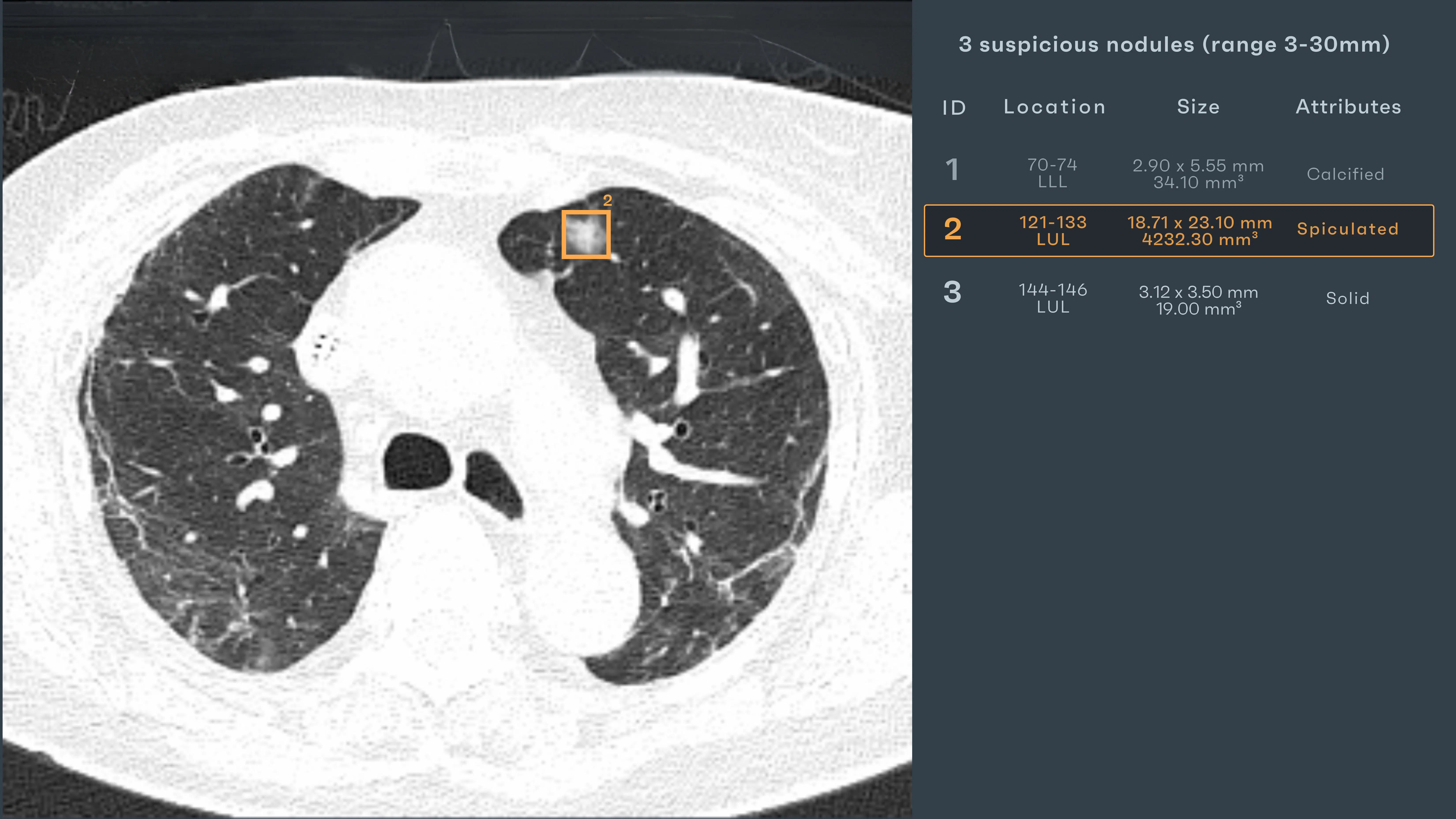
His scan had come three months earlier—baseline. Three nodules, one concerning: spiculated, 23.1 mm, 4232 mm³. It had been measured, reported, handed off. Now it was back.
The image looked familiar. That made it harder. Because this isn’t about finding something new. It’s about deciding whether what was once large is now less. Whether it shrank enough. Whether it still matters. And doing that not just with your eyes, but with criteria: RECIST, volumes, percentages, definitions so rigid they bend perception around them.
The radiologist pulled up the prior. Mentally recalibrated. Had it really shrunk? By how much? Enough? Because in cancer follow-up, the question isn’t just whether it’s smaller —it’s whether it means something.
AI Interaction
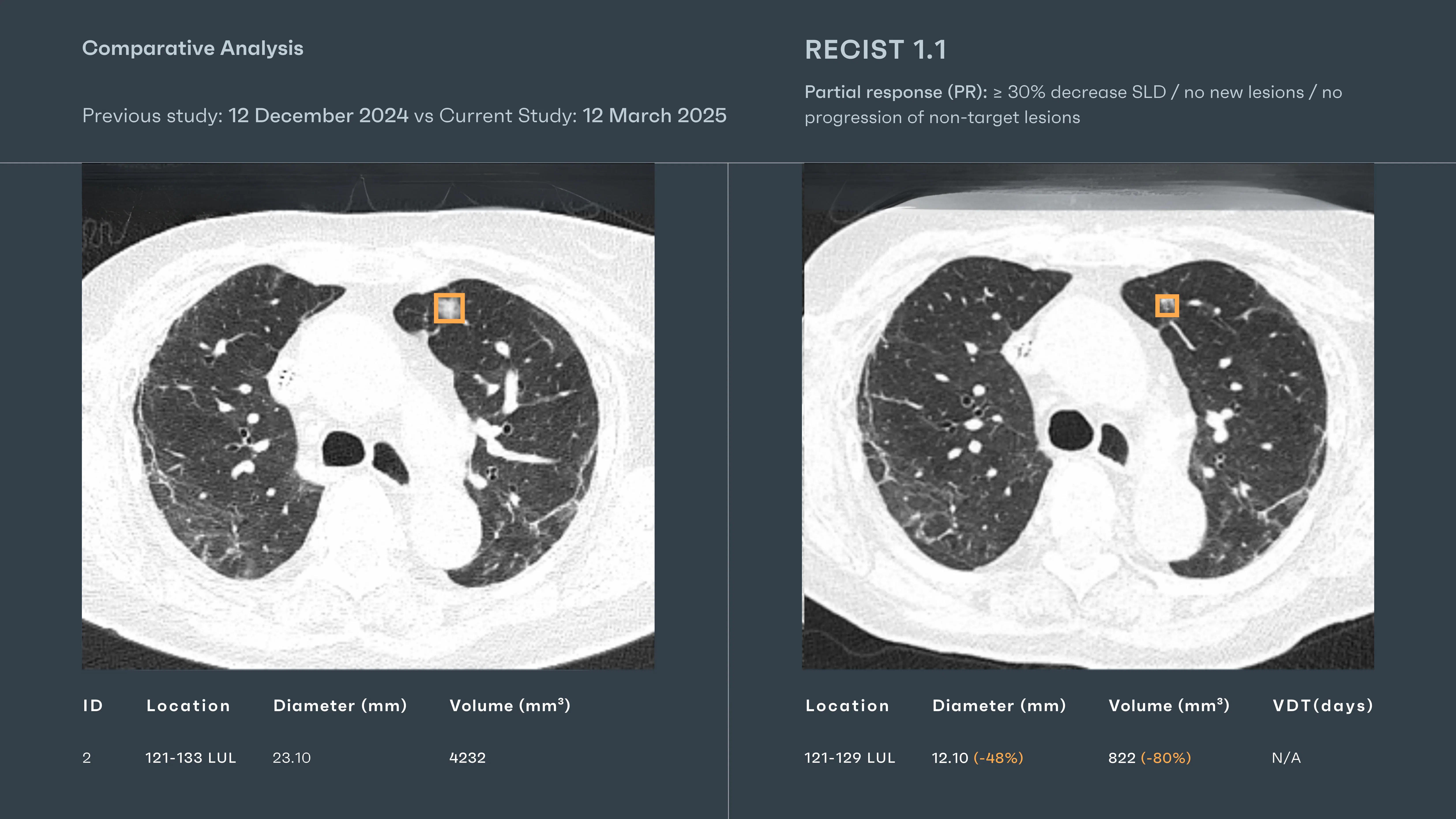
Rayscape ran its longitudinal module in the background. Side-by-side comparisons surfaced automatically:
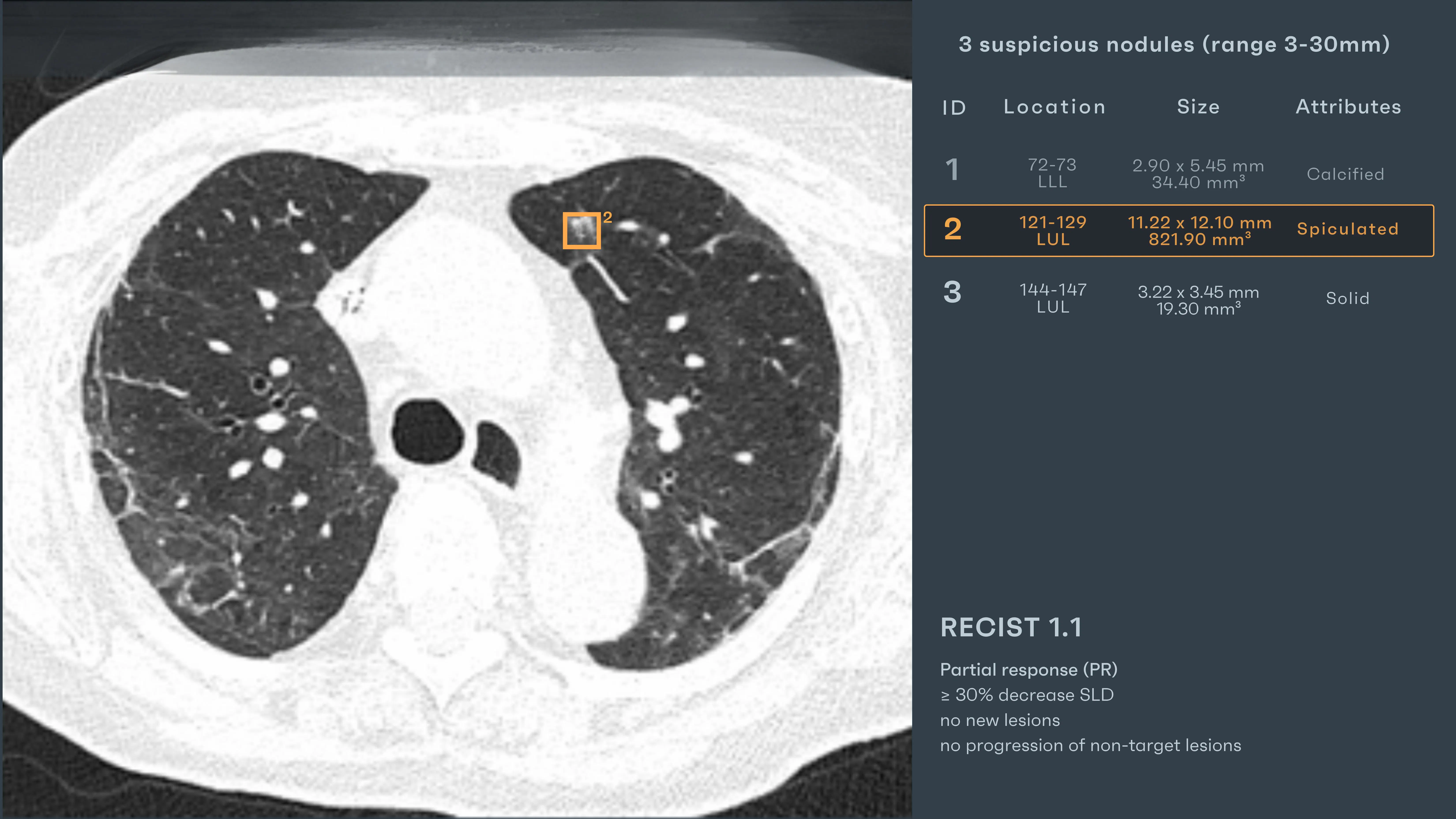
- Target Nodule – Previously 23.1 mm, now measured at 12.1 mm, volume 832 mm³
- AI-generated RECIST 1.1 category – Partial Response (PR) ≥30% decrease in SLD
- No new lesions detected
- No progression of non-target lesions
It measured and structured what the eye already suspected, and what the clinical team needed quantified. There was no need for scrolling back and forth, no manual segmentations. Just numbers—clear, stable, repeatable.
The decision wasn’t easy but the data was. The path was clearer and that, sometimes, is what counts most.
#ONCOLOGY CASE 2: A dot stable in time
Patient: Female, 65 years old
Setting: Oncology Follow-Up
Clinical Presentation: Monitoring known lesion under treatment
Imaging: Baseline CT (January) and Follow-up CT (March) - 2025
Clinical Context
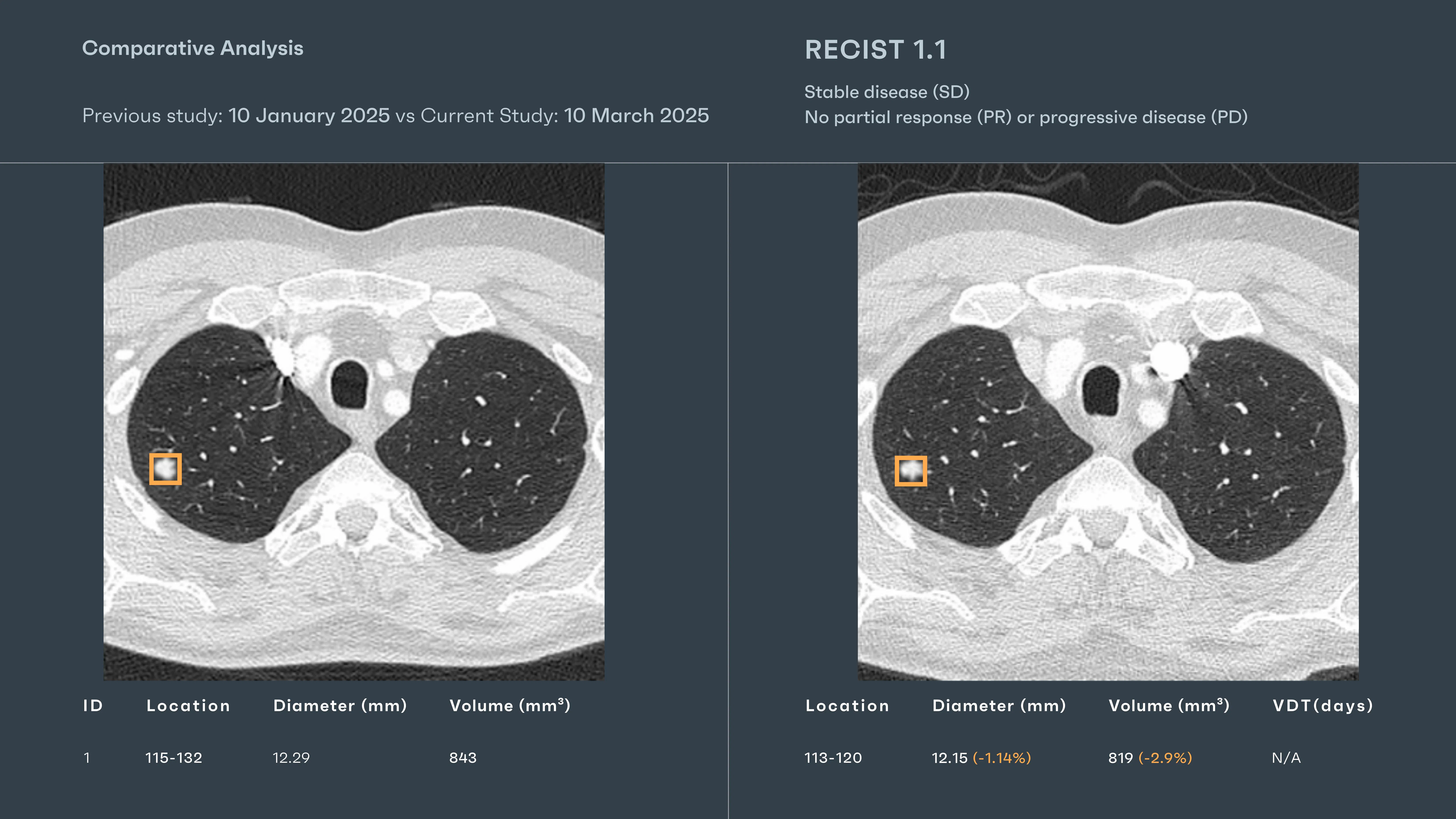
The follow-up folder said “no new symptoms.” That’s often worse than having some. Because in these cases, stability is not peace—it’s ambiguity. The nodule was first measured by AI in January: 12.29 mm, 843 mm³. Nothing dramatic, but enough to follow. Enough to hope it might shrink.
By March, the patient had already seen two oncologists, three infusion cycles, and countless side glances at words like “response.” But when the scan landed in the radiologist’s queue, there were no answers—only numbers, angles, and the quiet expectation to compare.
The cursor lingered. The mind wandered—was it windowing? Was it breath hold? Would someone misread this as progress? This is where fatigue becomes dangerous—not because something is missed, but because something is misunderstood.
AI Interaction
Rayscape processed both studies in the background—same algorithm, same structure, same baseline logic.
Target Lesion
- Previous: 12.29 mm / 843 mm³
- Current: 12.15 mm / 819 mm³
- Volume change: -2.9%
AI-assigned RECIST 1.1 category: Stable Disease (SD)
- No PR (≥30% decrease)
- No PD (≥20% increase)
- No new lesions
The overlays confirmed alignment. No growth. No shrinkage that mattered. Just...stillness. It measured. Giving the radiologist permission to accept what was—and not imagine more than that.
Outcome
The report was clear: “Nodule in shows minimal volumetric change (–2.9%) compared to prior. AI-calculated RECIST 1.1 classification: Stable Disease (SD). Recommend continued imaging surveillance.” No decisions were rushed. Sometimes, a dot stays where it is and that, too, is something to know.
#ONCOLOGY CASE 3:Beneath the numbers
Patient: Male, 70 years old
Setting: Oncology Follow-Up
Clinical Presentation: Surveillance CT for known solitary pulmonary nodule
Imaging: Baseline CT (October 2022) + Follow-up CT (October 2023)
Clinical Context
This scan came no symptoms, no urgent note. Just a quiet slot in a familiar name’s folder, tucked between a spine re-check and a post-op brain MRI. The last scan had been a year ago.
In this field, growth is its own language—subtle, statistical, and devastatingly clear. The burden isn’t just seeing the difference. It is being sure the difference meant something. The radiologist zoomed in. Scrolled. Adjusted windowing.
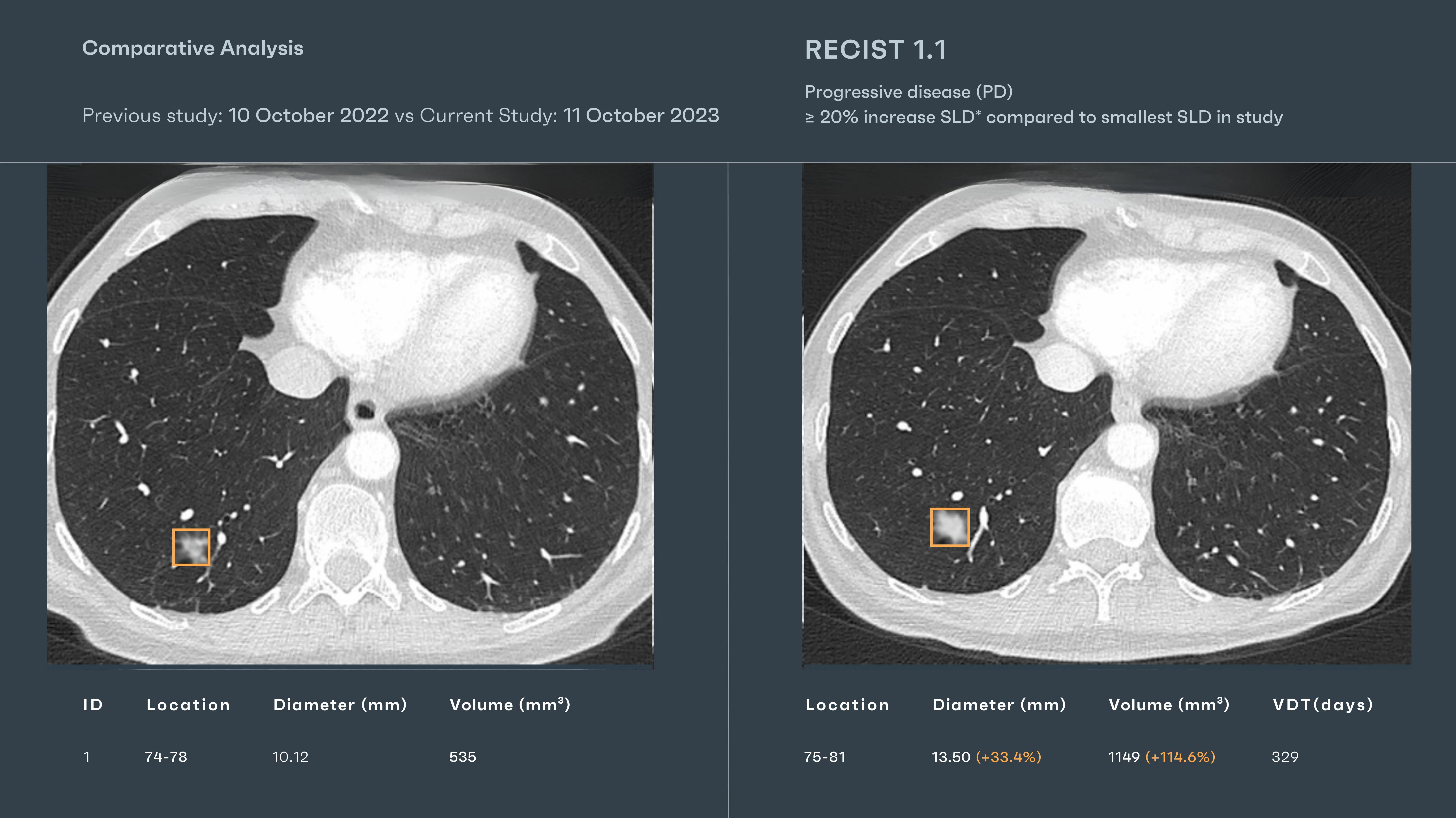
AI Interaction
Rayscape ran its timeline logic in the background. No urgency, no alarm—just math, clean and unblinking:
Target Nodule
- October 2022: 10.12 mm / 535 mm³
- October 2023: 13.50 mm (+33.4%) / 1149 mm³ (+114.6%)
- Volume Doubling Time (VDT): 329 days
AI-generated RECIST 1.1 classification: Progressive Disease (PD)
- ≥20% increase in sum of longest diameters compared to nadir
- Growth confirmed across both dimensions
This was a malignancy moving forward. The treatment wasn’t working.
Outcome
The report stated it plainly: “Target lesion shows increase over 12 months. RECIST 1.1 classification: Progressive Disease (PD).” The overlay didn’t dramatize—it simply stood, precise and annotated with volume trajectories visualized and metrics structured.
Discussions
Radiologists aren’t tireless or immune to overload. Behind every lung CT report is a reader juggling timelines, growth patterns, and nuanced findings—while the next scan waits in the queue. The pathology wasn’t hidden. It lived in ambiguity: a borderline nodule, a subtle ground-glass opacity, a shift just below the threshold of alarm.
The AI didn’t “catch” what the radiologist missed—it offered clarity before the fog could form. It positioned the findings within the frame, allowing the mind to move faster, the pattern to resolve more sharply. And this is the quiet revolution: not replacement, but reinforcement. Not autonomy, but augmentation. Rayscape acted not as a second opinion, but as a second set of steady hands in a moment where one pair may not be enough.
The future of lung CT is collaborative. Where human intuition meets algorithmic precision. Where a bounding box becomes not a conclusion, but a question, well-posed, in just the right place.
Built to fit your workflow
For IT teams and PACS administrators, implementation is often a key concern. Rayscape is designed to fit into your existing systems:
- Compatibility with major PACS platforms and cloud environments
- Vendor agnostic: trained to work on every DICOM image, no matter the vendor of the machine or the protocol
- Flexible deployment: on-premise, hybrid, or cloud-based
- Various outputs: SC, GSPS, SR
- Fast onboarding and secure data management protocols (HIPAA / GDPR compliant)
- Available on all major marketplaces (Deepc, Blackford, Carpl) or through direct sale
- We work closely with your radiology and IT departments to ensure seamless rollout with minimal disruption.
Let’s Talk
Your team makes life-saving decisions every day. Rayscape is here to support you in those critical moments—bringing clarity, speed, and confidence to outpatient imaging.
👉 Contact us and learn how we can empower your radiology team—when it matters most.

Discover how Rayscape works
integrate Rayscape AI into
your hospital's infrastructure
and improve both your
daily workflow and patients' experience.
