

Lung CT AI for Routine Clinical Follow-Up
Lung cancer rarely screams. It develops in silence, growing behind ribs and over years—quietly outpacing symptoms, eluding suspicion.
Many patients come to monitoring often sideways: after an incidental finding or through scattered follow-ups across time and institutions. Even when the scan is performed correctly, the context may be lacking. A missing prior. A half-completed note. A vague referral. The radiologist is left to reconstruct meaning from pixels—and must do so with speed, precision, and consistency.
Next, you’ll explore cases where Rayscape’s AI supported radiologists not with conclusions, but with structured comparisons. Highlighting subtle growth. Reassuring stasis.
The reality of real-world lung CT follow-up
Even when the scan quality is high, the risks remain:
- Small nodules may evolve incrementally—just enough to fall below suspicion
- Prior scans may not be available or properly aligned for comparison
- Time constraints push radiologists to triage findings rather than track trends
- Judgment fatigue can dull the recognition of change over time
How AI makes a difference
- Detects nodules and growth patterns across timepoints with volumetric precision
- Flags size and volume changes using consistent measurement logic
- Automatically aligns and compares with prior studies
- Offers transparent overlays and metrics without interrupting the reading workflow
Rayscape doesn’t decide what’s important. It gives the radiologist more tools to decide well. Where attention must stretch across months and sometimes years, AI helps ensure that even the quiet changes get a second look.
Below, you’ll encounter real clinical follow-up cases—usually not from screening programs, but from routine evaluations,. Each scenario shows how Rayscape’s AI supported radiologists in confirming:
- Nodule stability
- Interval growth
- Volume-based risk categorization
Each case is anonymized and presented with AI overlays and structured measurements as they appeared in daily clinical use. No simulations. No hypothetical projections. Just the real work of noticing change—before it matters too late.
#FOLLOW-UP CASE 1: The change was everything
Patient: Male, 66 years old
Setting: Outpatient Pulmonary Follow-Up
Clinical Presentation: Asymptomatic, history of prior screening CT
Imaging: Chest CT
Clinical Context
He didn’t feel any different.
No new symptoms. No new concerns. Just a follow-up, scheduled like a calendar reminder.
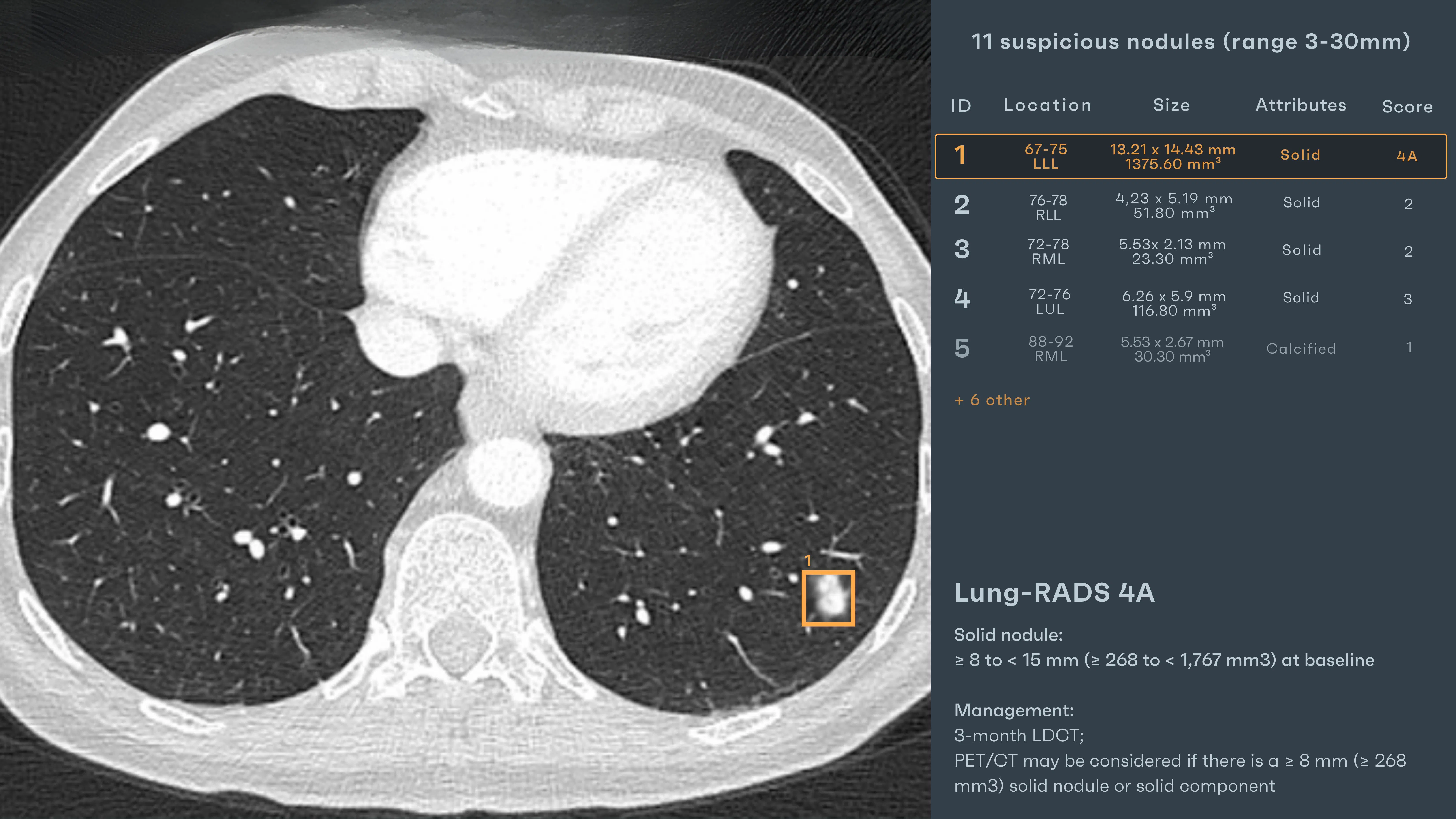
His previous CT had flagged a solid nodule in the lower left lobe—moderately sized, well-defined, suspicious enough to earn a Lung-RADS 4A. But also, not aggressive.
Not fast. Not, yet, anything more.
That’s the tension in follow-up care. The urgency doesn’t shout—it hums. A quiet pressure to decide what has or hasn’t changed. And under the weight of dozens of comparative reads a day, even that simple question— “Has this grown?”—can become surprisingly complex.
As the follow-up CT loaded, Rayscape automatically retrieved the prior scan. Without
clicks, without toggles, the overlay appeared—layered across both timepoints.
AI Interaction
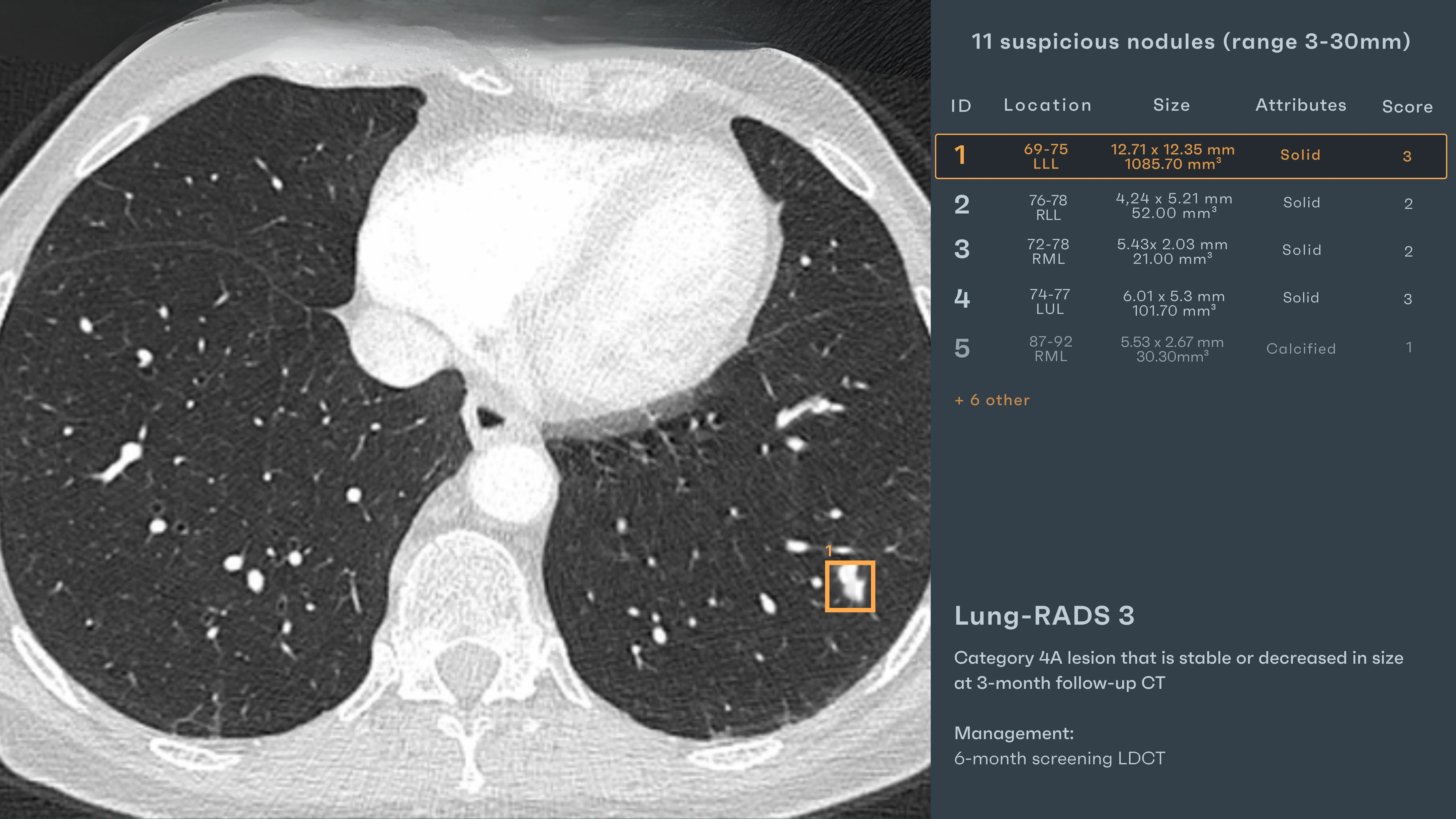
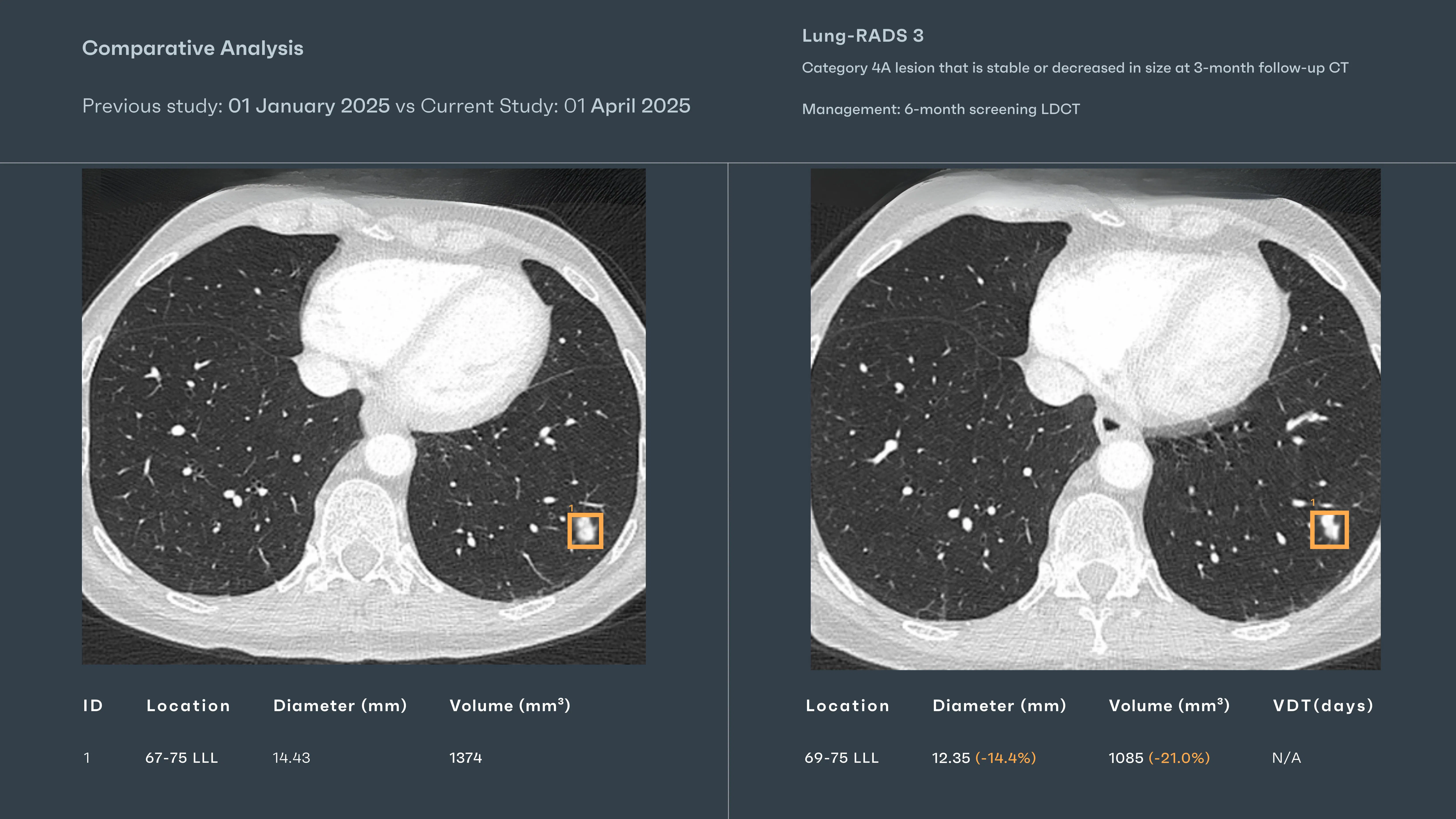
- “Nodule (LLL): 12.35 mm, 1085.7 mm³” – A precise box around the known lesion, now smaller than before.
- “Δ Volume: -21.0% | Δ Diameter: -14.4%” – Annotated inline, the AI flagged this as a significant reduction, consistent with post-inflammatory resolution or treated response.
- “Lung-RADS 3”– The system displayed the updated risk category automatically, downgrading the score in light of the interval change.
The overlays didn’t override judgment—but they reframed focus. The radiologist, who had reviewed three similar scans earlier that day, paused. The volume drop wasn’t obvious on first glance. But now it was documented, measured, contextualized. The report noted stability with interval reduction. Recommend continued 6-month follow-up.
#FOLLOW-UP CASE 2: The shift you could miss
Patient: Male, 59 years old
Setting: Routine Annual CT Follow-Up
Clinical Presentation: No symptoms, non-smoker, prior incidental pulmonary nodule
Imaging: Chest CT
Clinical Context
It’s easy to think of growth as something obvious—fast, expanding, loud. But in lung imaging, change is rarely theatrical. It happens slowly, hidden in decimal points and ratios. Not in a headline, but in a footnote.
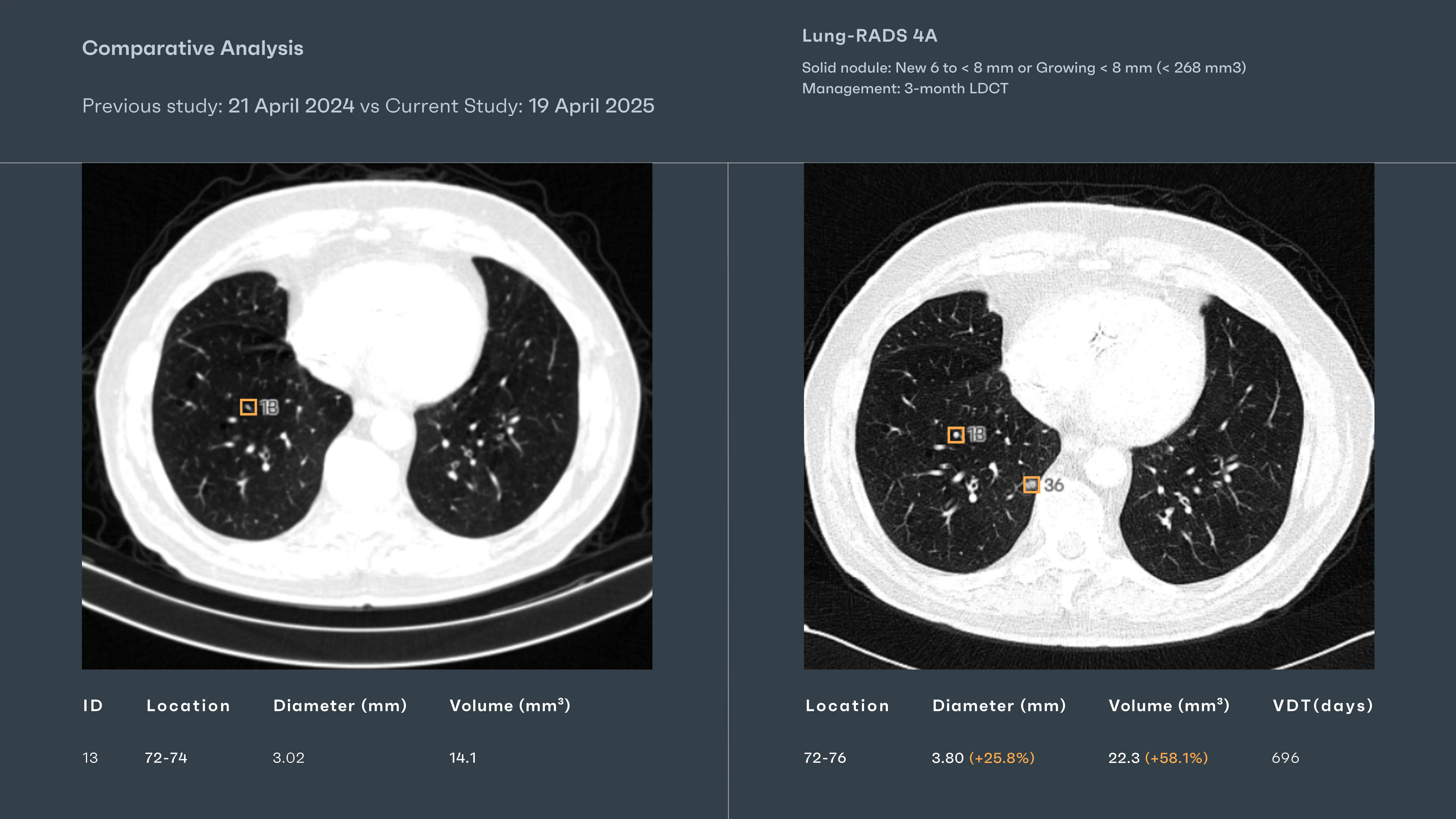
He was here for an annual check. His prior scan, one year earlier, had shown a small, benign-appearing nodule: 3.02 mm, 14.1 mm³. Watchlisted, but not alarming. The kind of case that floats near the bottom of the mental stack.
The 2025 scan came through on a busy morning. The radiologist skimmed the series: no effusion, no new consolidation, no dramatic change. But Rayscape’s AI didn’t skim. It compared. It measured. It noticed.
AI Interaction
Rayscape automatically aligned the current and prior CTs. Across the screen, a structured overlay locked in on the known nodule:
“Solid Nodule: 3.80 mm, 22.3 mm³” - A subtle, tight box surrounded the lesion. Not striking by sight—but the annotation was.
“Δ Volume: +58.1% | Δ Diameter: +25.8%”
“Volume Doubling Time: 696 days”
The radiologist leaned in. Without AI, it might have passed for stable. But here, side-by-side, the shift was visible.
Outcome
The report was updated with structured measurements and classification: “Interval growth of previously noted right lower lobe solid nodule from 3.02 mm (14.1 mm³) to 3.80 mm (22.3 mm³), representing a 58.1% volume increase. Recommend 3-month low-dose CT follow-up.” No immediate alarm but the case moved off the ‘routine’ pile. Subtle growth had been seen—and this is enough to matter.
#FOLLOW-UP CASE 3: Stillness is also a sign
Patient: Male, 63 years old
Setting: Longitudinal CT Surveillance
Clinical Presentation: No symptoms, nodule flagged in prior scan
Imaging: Chest CT
Clinical Context
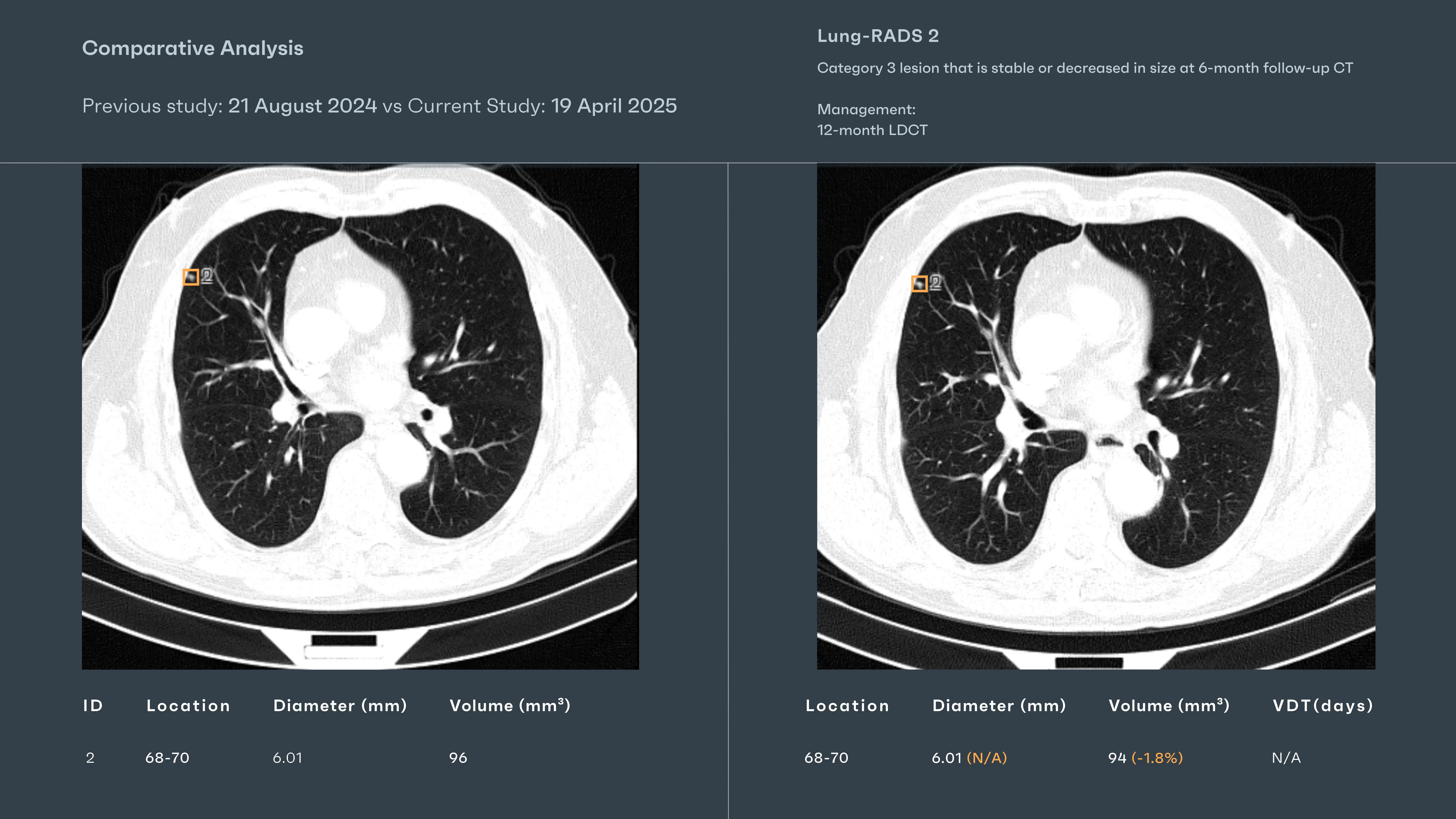
Some stories unfold quickly. Others linger—quiet, uneventful, steady in their ambiguity. The patient was part of a routine follow-up program. A nodule had been flagged the year before: 6.01 mm, 96 mm³, solid and well-circumscribed. Enough to warrant a closer look. Not enough to justify alarm.
This scan was his return. Just another datapoint in the slow watchfulness that defines screening programs everywhere.
AI Interaction
Rayscape ran the comparison the moment the study was loaded. The nodule, now familiar, was immediately highlighted.
- “Solid Nodule: 6.01 mm, 94 mm³” – A slight drop in volume, measured precisely.
- “Δ Volume: -1.8% | Δ Diameter: 0%” – A subtle shift, but not significant.
The AI didn’t flag urgency. But it framed continuity. In a stack of dozens of follow-up reads, it quietly said: this one is holding still. The report recommend continued surveillance with 12-month low-dose CT. No headlines and no change in course, but in the world of lung cancer screening, sometimes the most powerful result is the absence of motion.
Discussions
Radiologists aren’t tireless or immune to overload. Behind every lung CT report is a reader juggling timelines, growth patterns, and nuanced findings—while the next scan waits in the queue. The pathology wasn’t hidden. It lived in ambiguity: a borderline nodule, a subtle ground-glass opacity, a shift just below the threshold of alarm.
The AI didn’t “catch” what the radiologist missed—it offered clarity before the fog could form. It positioned the findings within the frame, allowing the mind to move faster, the pattern to resolve more sharply. And this is the quiet revolution: not replacement, but reinforcement. Not autonomy, but augmentation. Rayscape acted not as a second opinion, but as a second set of steady hands in a moment where one pair may not be enough.
The future of lung CT is collaborative. Where human intuition meets algorithmic precision. Where a bounding box becomes not a conclusion, but a question, well-posed, in just the right place.
Built to fit your workflow
For IT teams and PACS administrators, implementation is often a key concern. Rayscape is designed to fit into your existing systems:
- Compatibility with major PACS platforms and cloud environments
- Vendor agnostic: trained to work on every DICOM image, no matter the vendor of the machine or the protocol
- Flexible deployment: on-premise, hybrid, or cloud-based
- Various outputs: SC, GSPS, SR
- Fast onboarding and secure data management protocols (HIPAA / GDPR compliant)
- Available on all major marketplaces (Deepc, Blackford, Carpl) or through direct sale
- We work closely with your radiology and IT departments to ensure seamless rollout with minimal disruption.
Let’s Talk
Your team makes life-saving decisions every day. Rayscape is here to support you in those critical moments—bringing clarity, speed, and confidence to outpatient imaging.
👉 Contact us and learn how we can empower your radiology team—when it matters most.


Discover how Rayscape works
integrate Rayscape AI into
your hospital's infrastructure
and improve both your
daily workflow and patients' experience.
