

AI for Radiologists in ICU Chest X-rays
The Intensive Care Unit is a place of constant vigilance. Patients are monitored by the second, managed by multi-disciplinary teams, and treated within windows that leave little room for delay. Amid ventilators, vasopressors, and sterile drapes, imaging plays a critical—but often quiet—role in steering care.
But here too, the story is familiar. Imaging volumes grow. Interpretations must be rapid, reliable, and revisited. Radiologists read in the context of evolving pathology, limited patient positioning, and complex clinical backgrounds. The margin for uncertainty narrows—while the demand for clarity expands.
Next you’ll explore clinical scenarios where Rayscape’s AI doesn’t take charge, but takes its place—alongside the radiologist, in the layered, high-stakes reality of critical care. A perceptive observer, it highlights what progression may mask, tracks changes across days, and offers visual precision in moments where time moves faster than attention.
What is Rayscape?
Rayscape is a radiology AI company focused on augmenting radiologists and clinical teams in their daily decision-making. Our technology assists in the interpretation of chest X-rays and lung CT scans, using large-scale medical image training to detect, measure, and monitor key findings.
Designed by radiologists, for radiologists, Rayscape is built with real-world workflow in mind—delivering support where it matters most: during on-call shifts, under pressure, and in the screening or monitoring of oncology patients—environments where speed and accuracy are critical.
Built to support, not replace- Rayscape assists the radiologist in seeing more, seeing earlier, and seeing clearly.
Saving time, saving lives - the role of AI in the ICU
In the ICU, chest imaging isn’t about incidental findings—it’s about survival. One that evolves hour by hour, scan by scan. Radiologists are often tasked with identifying minute changes that hint at worsening respiratory distress, infection, or cardiac instability.
Clinicians rely on fast reporting for life-threatening decisions. But real-world challenges persist:
- Portable X-rays with limited positioning and suboptimal contrast
- Devices and lines obscuring key findings
- Limited time for careful serial review across imaging studies
- Fatigue and volume can impact even the most experienced radiologist’s performance
Here’s how Rayscape makes a difference
- Support radiologists with instant AI-generated highlights that draw attention to overlooked or evolving findings
- Detects device placements, lung abnormalities, and pleural pathologies with visual overlays
- Ensure consistency in image analysis, regardless of time of day or workload
- Offer structured overlays and measurements, without disrupting the existing workflow
With Rayscape, radiologists can focus on what they do best—making life-saving decisions—while we help ensure nothing critical gets missed.
Proven clinical value
In the following, you’ll encounter real clinical cases from intensive care units where Rayscape supported the diagnostic process in critical patients—each one showcasing how AI has supported radiologists in detecting, confirming, and communicating urgent findings such as:
- Progressive pleural effusions
- Subtle pneumothoraces under pressure ventilation
- Lung infiltrates
- Lung consolidations
...and more.
Each case is anonymized and presented with AI overlays as they appeared in practice. These are not hypothetical scenarios—they are taken from daily workflows, under real clinical pressure.
#ICU CASE 1: The shadow that shifted overnight
Patient: Female, 82 years old
Setting: Intensive Care Unit
Clinical Presentation: Post-trauma monitoring, intubated, mild hypotension, increased oxygen demand overnight
Imaging: Portable Chest X-ray
Clinical Context
Cases don’t line up politely in the ICU. They arrive layered—stacked between stroke alerts, scheduled abdomens, emergent trauma, and the steady background hum of portables. ICU films often slip through in clusters, squeezed between the urgent and the routine, their filenames familiar but their urgency uncertain.
Fatigue works differently here. It doesn't rush in—it accumulates. The radiologist scrolls through lung fields while juggling temporal comparisons, line placements, surgical follow-ups, and the haunting promise of missed context.
There’s a rhythm to ICU reads: quick but cautious, clinical notes half-finished, images partially rotated. And in that rhythm, there are blind spots. Not through neglect, but through volume. A fluid rim mistaken for artifact. A subtle opacity absorbed into routine. These aren’t misses—they’re the edges where human perception can blur under constant strain.
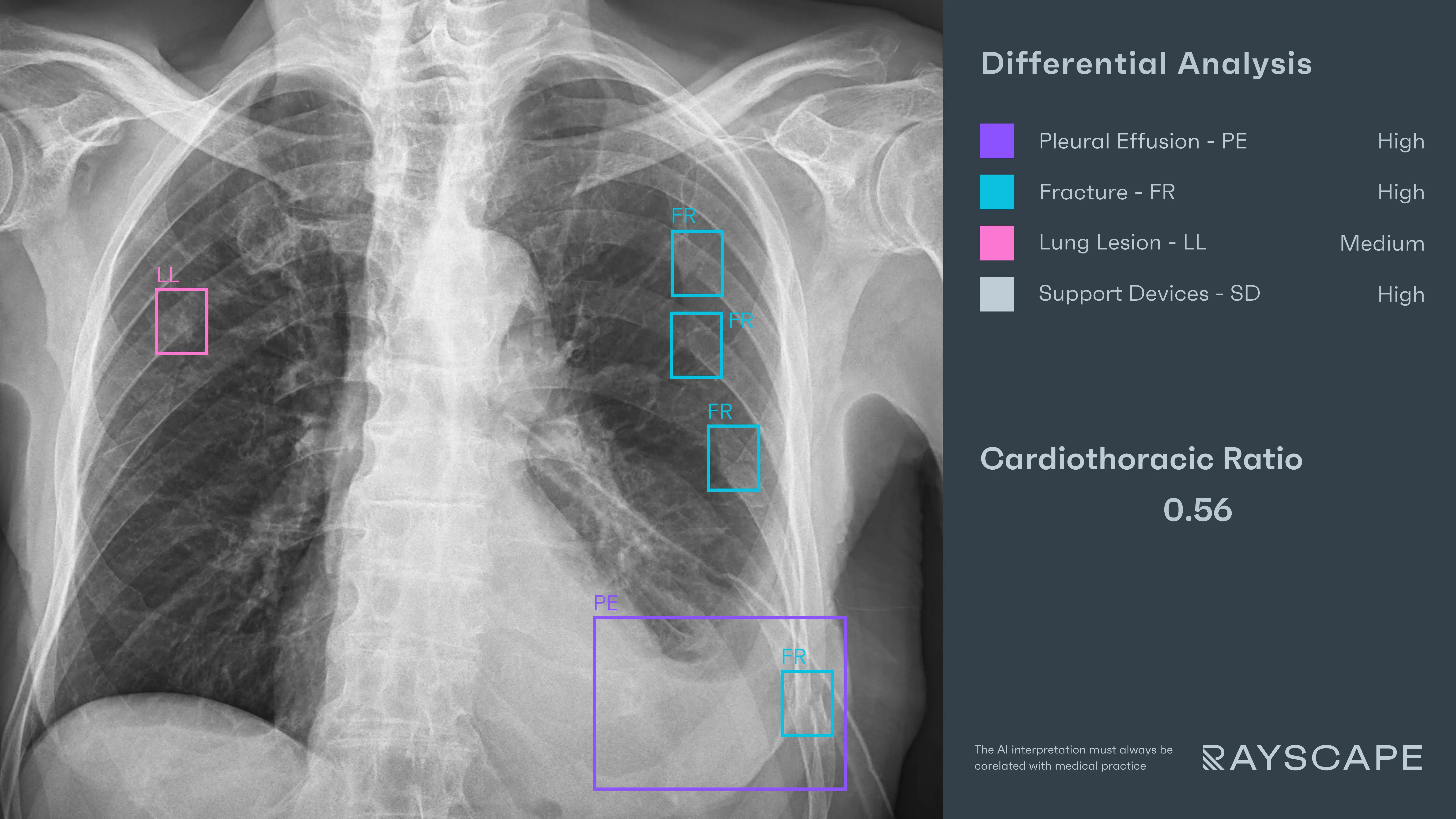
She was 82. Elderly, frail, brought in after a fall that left her with rib fractures on the left. She had been intubated for comfort and monitoring, and the clinical team was attentive. But overnight, her oxygen requirements crept upward. Not dramatically—but enough to ask for imaging. A chest X-ray was ordered before rounds.
AI Interaction
As the chest X-ray loads into the PACS viewer, the Rayscape AI assistant runs its analysis in parallel. Within seconds, its interface overlays multiple distinct bounding boxes:
- "Pleural Effusion" – A bounding box arced across the left lower hemithorax, highlighting a meniscus and rising fluid, moderate in volume.
- Left Rib Fractures" – Bounding boxes marked fractures on the left ribs, with visible cortical discontinuity and mild displacement.
- "Lung Lesion" – A second box captured a dense, irregular patch in the right lung—poorly defined, suggestive of consolidation, possibly infectious in origin.
The algorithm did not overstate its role. It didn’t diagnose or suggest urgency. It simply framed what needed framing—clean, consistent, quietly placed in the radiologist’s line of thought.
The radiologist paused. What at first seemed like a routine check suddenly carried weight: fluid on the left where there hadn’t been, and an emerging opacity on the right, sharp enough to demand follow-up. Subtle changes, but in the ICU, subtle doesn’t mean small.
These were not dramatic alarms. They were signposts—small, sharp gestures meant to guide attention to a possible infection.
Outcome
Thoracic ultrasound confirmed the effusion, and it was drained that afternoon. The right-sided opacity was clinically correlated and empiric antibiotics initiated. The patient’s respiratory status improved—course corrected before the drift became a spiral.
#ICU CASE 2: A cloud without air
Patient: Female, 76 years old
Setting: Intensive Care Unit
Clinical Presentation: Severe hemoptysis, desaturation, history of vasculitis
Imaging: Portable Chest X-ray
Clinical Context
The call came at 04:17. A note scribbled into the imaging system: “desaturation + hemoptysis overnight, rule out pneumonia.” One of the nurses had mentioned blood in the ETT filter.
She was 76. Known vasculitis. Recently transferred from internal medicine after sudden hypoxia. Sedated, intubated, PEEP rising. The team didn’t have time to wait for bronchoscopy—they needed a direction now. The X-ray arrived in the quiet space before morning rounds. And while the image itself was hazy, the urgency was not.
AI Interaction
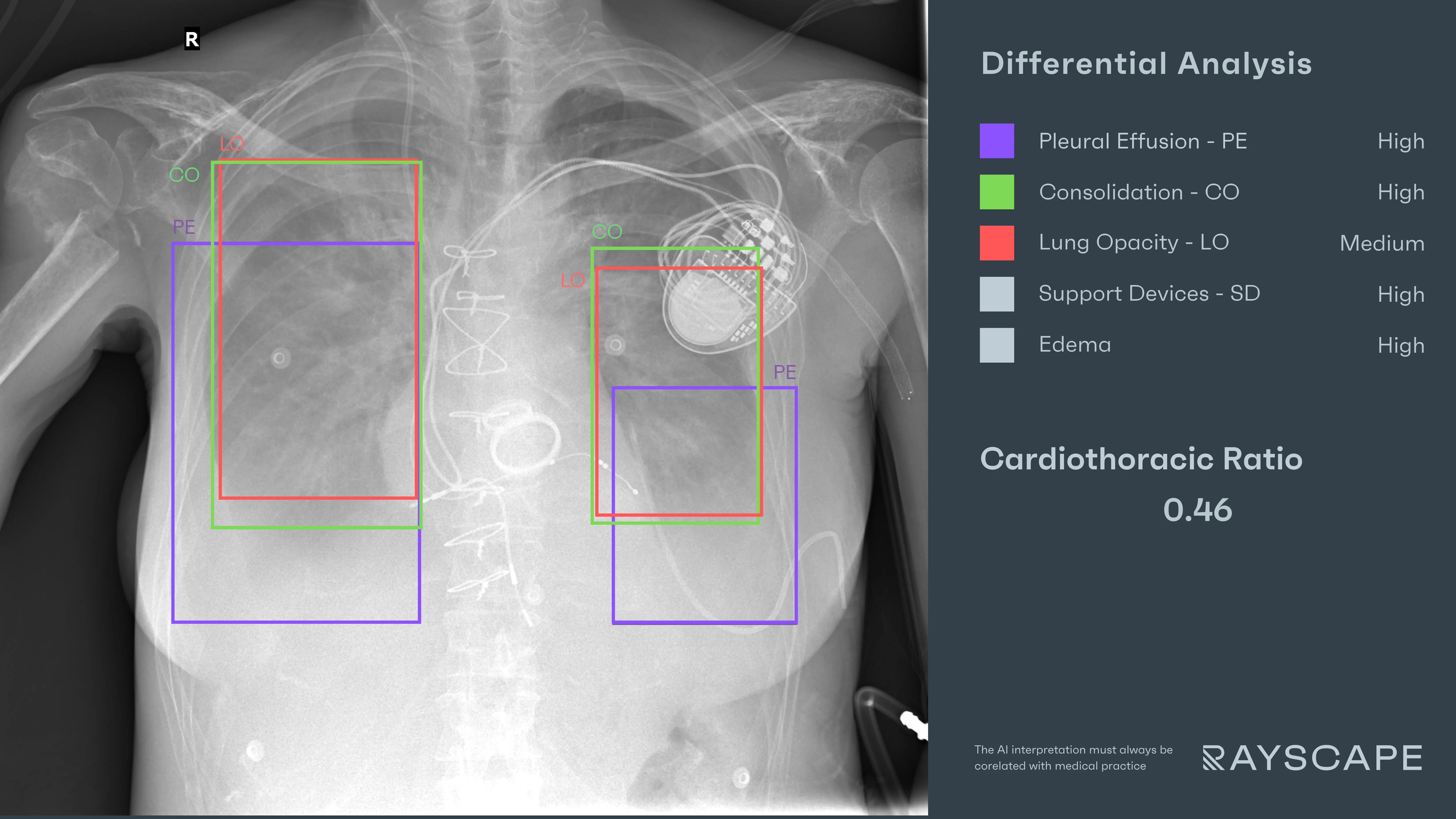
Rayscape’s AI rendered the image in silence, then placed its overlays across both lung fields:
- "Pleural Effusion" – Bilateral, faint, but moderate to high.
- "Lung Opacities" – Dense, symmetrical, bilateral shadows with air bronchograms lost in the grayscale—full-field consolidations, with no spared segments.
- "Consolidation" – Labeled again, across both lungs: diffuse, alveolar, suffocating in density.
The radiologist stared. No lobar pattern, no classic gradient, no infectious asymmetry. Just lungs filled with something that didn’t want to clear. Rayscape didn’t suggest a diagnosis—but in the shape of its boxes, the radiologist found clarity: this was not pneumonia.
She typed the words carefully: “Diffuse alveolar opacities concerning for alveolar hemorrhage in a known vasculitis patient. Recommend immediate bronchoscopy and supportive care.”
Outcome
Bronchoscopy confirmed blood in all segments. A diagnosis of diffuse alveolar hemorrhage was made. Immunosuppressive therapy escalated. Ventilator settings adjusted. She remained unstable—but now her story had a name.
#ICU CASE 3: The pressure within
Patient: Female, 85 years old
Setting: Intensive Care Unit
Clinical Presentation: Hypotension, history of atrial fibrillation, worsening dyspnea on non-invasive ventilation
Imaging: Portable Chest X-ray
Clinical Context
Not all collapses happen suddenly. Some happen over days—slowly, beneath a surface of mild hypoxia, rising BNP, vague malaise. She was 85, admitted two nights earlier for suspected pulmonary edema. But diuresis hadn't improved her condition. Now, her blood pressure was drifting downward, her O₂ demand creeping upward. The ICU team wanted to rule out pneumonia.
The X-ray came through during change of shift. The radiologist scanned it between scheduled CTs and a neuro follow-up. It looked familiar. And that was part of the danger.
AI Interaction
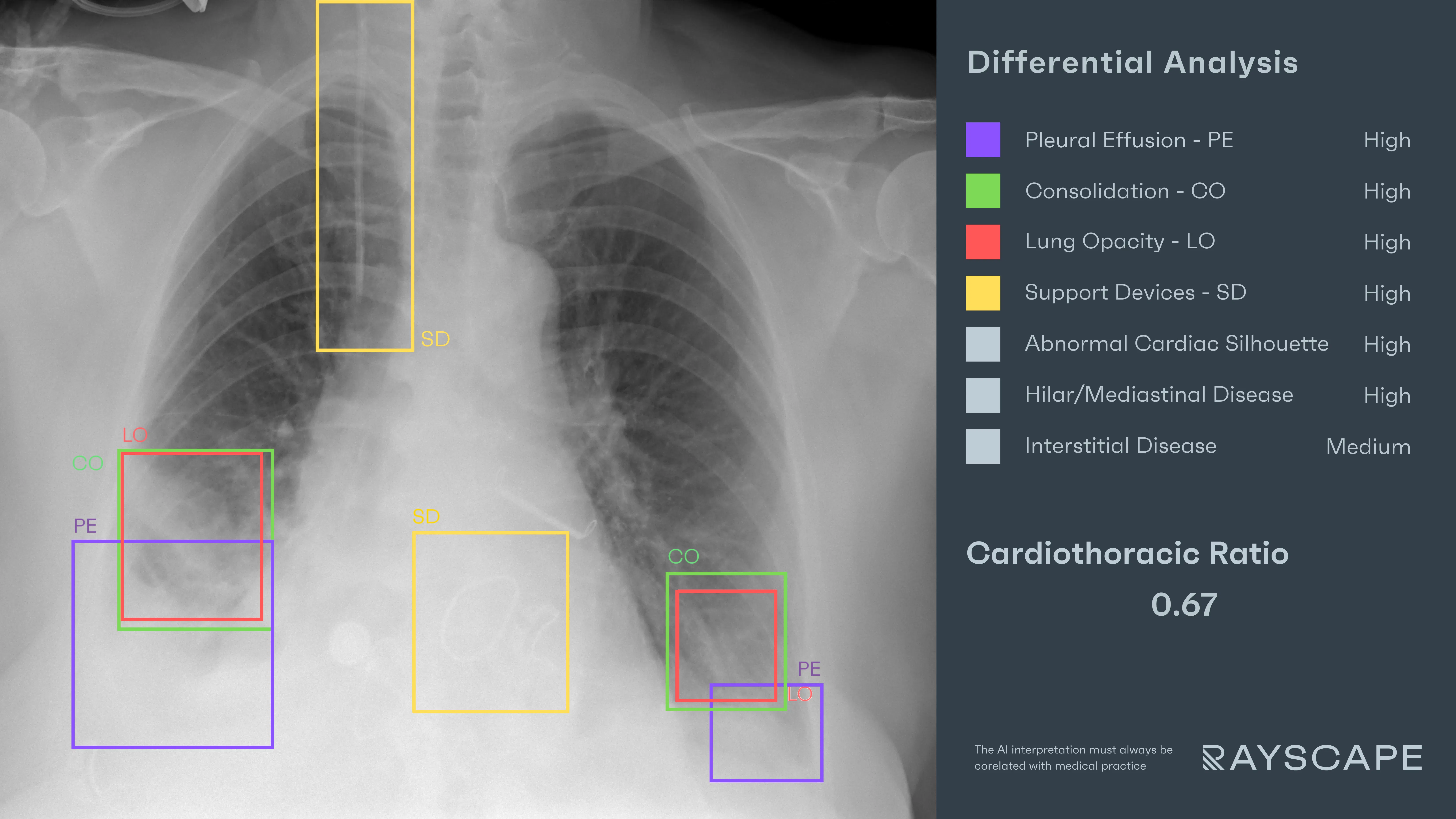
Rayscape stepped in—not to diagnose, but to steady the frame, to sort the noise from the signal.
- "Pleural Effusion" – Bilateral, small but persistent—clear crescents at the lung bases.
- "Consolidation" – Patchy opacities at the lower lobes, more prominent on the right—suggestive but not dominant.
- "Cardiothoracic Ratio: 0.67" – Elevated, indicating a visibly enlarged heart.
- "Abnormal Cardiac Silhouette" – The AI flagged the rounded, displaced – a widened contour.
The radiologist paused. This wasn’t classic heart failure. The cardiac silhouette looked stretched, not just large. There was a fullness behind the sternum—subtle, but not accidental. This wasn’t volume overload. This was a space problem.
She flagged it in the report: “Subtle findings concerning for pericardial effusion. Recommend echocardiographic correlation to assess for early tamponade physiology.”
Outcome
Echo was performed within the hour. It confirmed a moderate pericardial effusion with signs of hemodynamic compromise. Pericardiocentesis was arranged. Within two hours, her pressure had stabilized—and her breath returned.
Discussions
The ICU does not operate on urgency alone—it runs on the tension between stability and sudden collapse. And radiologists reading for the ICU know this well. They are not tireless, infallible, or untouched by the weight of it all. They work in the quiet corners of crisis—balancing subtle shifts in shadow against timelines that are anything but linear.
In these cases, the findings were not dramatic, but they were decisive. They did not scream—they accumulated. A contour displaced. A lung that filled not with air, but with silence. Rayscape didn’t uncover what was hidden—it clarified what had become hazy under the pressure of time and repetition.
This is where AI earns its place—not as a replacement, but as a safeguard. Not as the one who knows better, but as the one who remembers to look again. It adds structure to complexity. It steadies the gaze. It doesn’t change what is seen—it helps define when it matters most to see it.
The future of ICU imaging is not machines reading in place of humans. It is systems built to amplify clinical intuition, to catch the drift before it becomes a fall. It is a bounding box that doesn’t close the case—but opens the right question, at the right time.
Built to fit your workflow
For IT teams and PACS administrators, implementation is often a key concern. Rayscape is designed to fit into your existing systems:
- Compatibility with major PACS platforms and cloud environments
- Vendor agnostic: trained to work on every DICOM image, no matter the vendor of the machine or the protocol
- Flexible deployment: on-premise, hybrid, or cloud-based
- Various outputs: SC, GSPS, SR
- Fast onboarding and secure data management protocols (HIPAA / GDPR compliant)
- Available on all major marketplaces (Deepc, Blackford, Carpl) or through direct sale
- We work closely with your radiology and IT departments to ensure seamless rollout with minimal disruption.
Let’s Talk
Your team makes life-saving decisions every day. Rayscape is here to support you in those critical moments—bringing clarity, speed, and confidence to ICU imaging.
👉 Contact us and let’s talk about how we can empower your radiology team—when it matters most.

Discover how Rayscape works
integrate Rayscape AI into
your hospital's infrastructure
and improve both your
daily workflow and patients' experience.
