

AI Boosts Chest X-ray Reads in Outpatient Centers
Outpatient imaging operates at a different pace. There are no alarms, no sirens—only a quiet pressure that builds with every unclear clinical note and every half-answered question in the referral field.
Patients arrive for routine checks, pre-op evaluations, and persistent symptoms that haven’t resolved on their own. Some are anxious, some are asymptomatic. And radiologists are asked to make judgments based on a single scan, often with limited clinical context and no guarantee of follow-up.
When every case is a maybe, and every lung shadow could mean something—or nothing—what radiologists need isn’t a diagnosis delivered. It’s a system that helps sharpen their focus, frame ambiguity, and offer clarity without overstepping.
Next, you’ll explore outpatient cases where Rayscape’s AI works not as an authority, but as a lens—helping surface what matters, especially when there’s little else to go on. A perceptive assistant in a setting where the greatest challenge isn’t urgency, but uncertainty.
What is Rayscape?
Rayscape is a radiology AI company focused on augmenting radiologists and clinical teams in their daily decision-making. Our technology assists in the interpretation of chest X-rays and lung CT scans, using large-scale medical image training to detect, measure, and monitor key findings.
Designed by radiologists, for radiologists, Rayscape acts as a consistent second set of eyes. For outpatient clinics, it is helping identify incidental but actionable findings, surfacing missed abnormalities, and supporting early detection.
Built to support, not replace- Rayscape assists the radiologist in seeing more, seeing earlier, and seeing clearly.
Seeing sooner, acting faster- AI's impact in the Outpatient Settings
Unlike in the ER or ICU, outpatient imaging is rarely driven by crisis. But that doesn’t mean the stakes are lower. Many serious diseases—lung cancer, TB, heart failure—first present subtly, and often during a routine outpatient workup.
And yet:
- Clinical histories are often vague or missing
- Incidental findings risk being overlooked or dismissed as insignificant
- High volume can impact even the most experienced radiologist’s performance
Here’s how Rayscape makes a difference
- Draws attention to overlooked, incidental, or unexpected findings in low-acuity settings
- Helps surface subtle but actionable pathology—even in cases flagged as routine
- Supports early detection of abnormalities that may otherwise go unreported
- Delivers consistent analysis in high-volume, low-context imaging environments
With Rayscape, radiologists gain a quiet partner: one that helps ensure that even a routine exam gets the full depth of clinical attention—without slowing the workflow that outpatient centers depend on.
Proven clinical value
In the following, you’ll find clinical cases drawn from outpatient imaging centers—each one reflecting how Rayscape contributed to the identification and reporting of findings that could have easily gone unnoticed.
These include:
- Incidental pulmonary nodules
- Subtle pleural changes
- Early-stage consolidations
- Cardiomegaly
...and more.
Each case is anonymized and presented with AI overlays as they appeared in practice—under real outpatient conditions, in real radiologist workflows.
#OUTPATIENT CASE 1: The shadow beside the heart
Patient: Female, 67 years old
Setting: Outpatient Imaging Center
Clinical Presentation: Referred for persistent dry cough, no prior imaging available
Imaging: Chest X-ray
Clinical Context
There was no urgency attached to the order. Just a checkbox: “chronic cough.” No lab results. No auscultation notes. No smoking history. Just a time slot in the late afternoon, tucked between a post-op shoulder and a pre-op pelvis.
That’s the tempo in the outpatient center. The pressure doesn’t come from chaos—it comes from routine. From reading too many normal scans in a row, until they start to blur into a single visual rhythm. And in that rhythm, true pathology becomes harder to hear. Not because it hides, but because it’s surrounded by silence.
The cases arrive quietly, without triage urgency or flashing vitals. But for the radiologist, that silence becomes a different kind of risk. The kind that asks: “What if this is the only image she gets?”
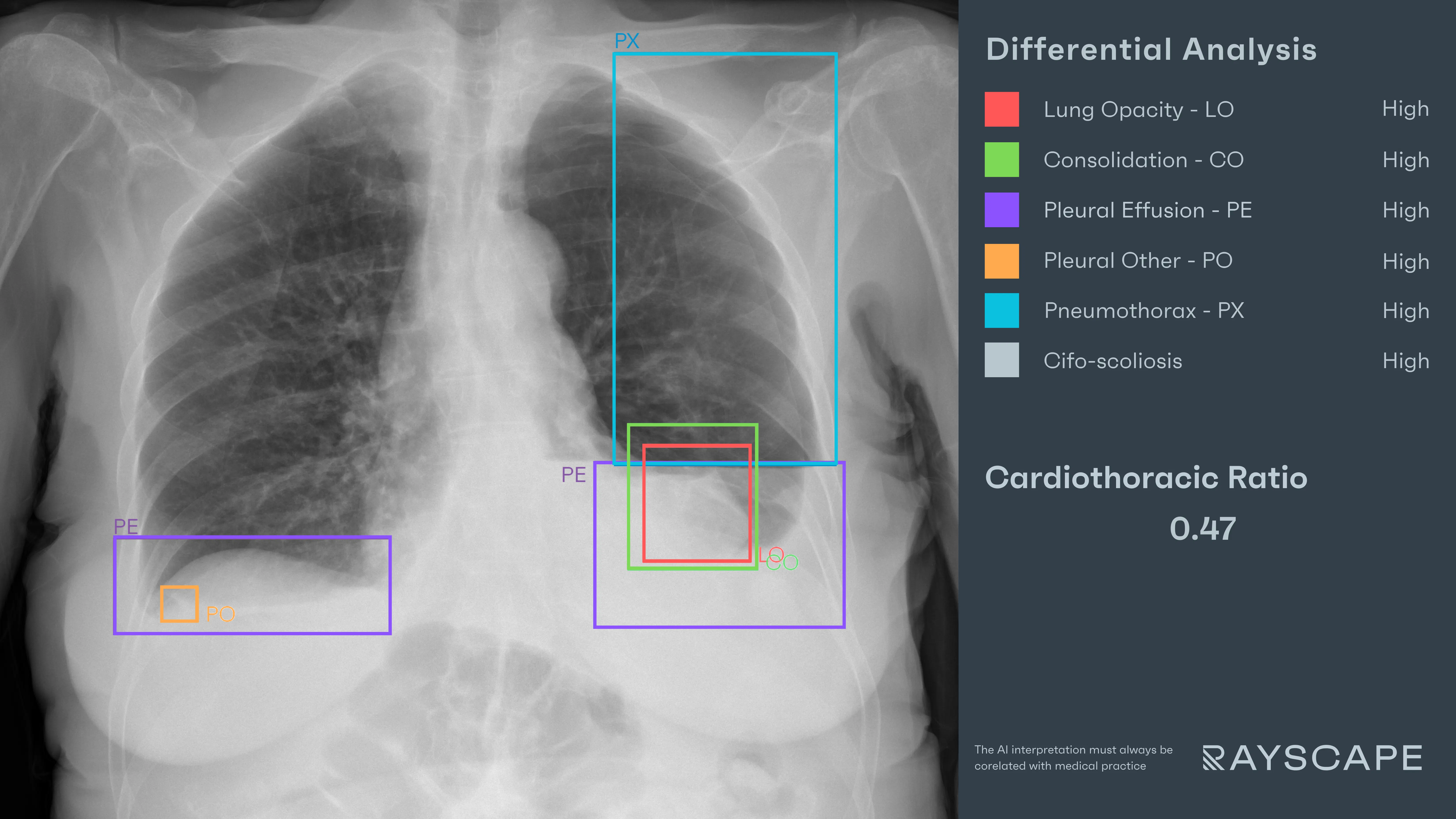
So the X-ray appears, like so many others. A slightly rotated view. The spine curved rightward. Ribs uneven in their spacing. Nothing dramatic—but something... off.
AI Interaction
As the chest X-ray loads into the PACS viewer, the Rayscape AI assistant runs its analysis in parallel. And in a moment, the findings began to surface:
- "Lung Opacity" – A bounding box hovered near the left heart border, identifying a soft density creeping toward the hilum. It was low, smooth, and easily mistaken for silhouette.
- "Consolidation" – The same area, now highlighted again, thicker, denser—suggesting true left lower lobe consolidation with possible perihilar involvement.
- "Pleural Effusion" – Bilateral, faint but definite. Thin fluid layers resting at the bases—more pronounced on the left.
- "Pneumothorax" – A subtle pleural line in the left upper zone. Easy to miss, delicate as a thread. But absent lung markings made the message clear.
- "Cifo-Scoliosis" – Finally, a gentle arc traced along the thoracic spine—moderate thoracic curvature, shifting both anatomy and expectations.
The radiologist leaned in. The curvature had drawn the mediastinum. The consolidation had blended into the heart. The effusion blurred the costophrenic edge. And the pneumothorax—small, yes—but not imaginary.
None of these findings were individually urgent. But together, they told a story far more complex than “chronic cough.”
Outcome
The report was issued with urgency revised. “Left lower lobe consolidation with underlying effusion and subtle pneumothorax. Suspicion of early parapneumonic complication. Recommend clinical follow-up and CT correlation.”
The referring physician called back within the hour. The patient was contacted and brought in for reassessment. A contrast-enhanced CT performed the next day revealed an evolving loculated pleural effusion with air-fluid levels, suspicious for early empyema, and areas of segmental necrosis within the left lower lobe.
Antibiotics were escalated, and interventional pulmonology was consulted for drainage. What began as a routine outpatient image quietly diverted the course of a more serious pathology.
#OUTPATIENT CASE 2: The nodule in the noise
Patient: Male, 58 years old
Setting: Outpatient Imaging Center
Clinical Presentation: Persistent cough, occasional wheezing, no prior chest imaging available
Imaging: Chest X-ray
Clinical Context
Some patients arrive at the outpatient center not for answers, but for reassurance. “It’s probably nothing.” “Just seasonal.” “My GP wanted to be thorough.”
This was one of those cases. A man in his late fifties, dressed like he had come from work. No weight loss. No fever. Just a cough that hadn’t left after the flu. The referral said, simply: “chronic cough – r/o pneumonia.”
The radiologist had already read four normal scans that hour. Shoulders, sinuses, a lateral knee. When the lungs finally appeared on screen, they looked routine enough—at first. Nothing urgent. Nothing screaming. But not nothing.
AI Interaction
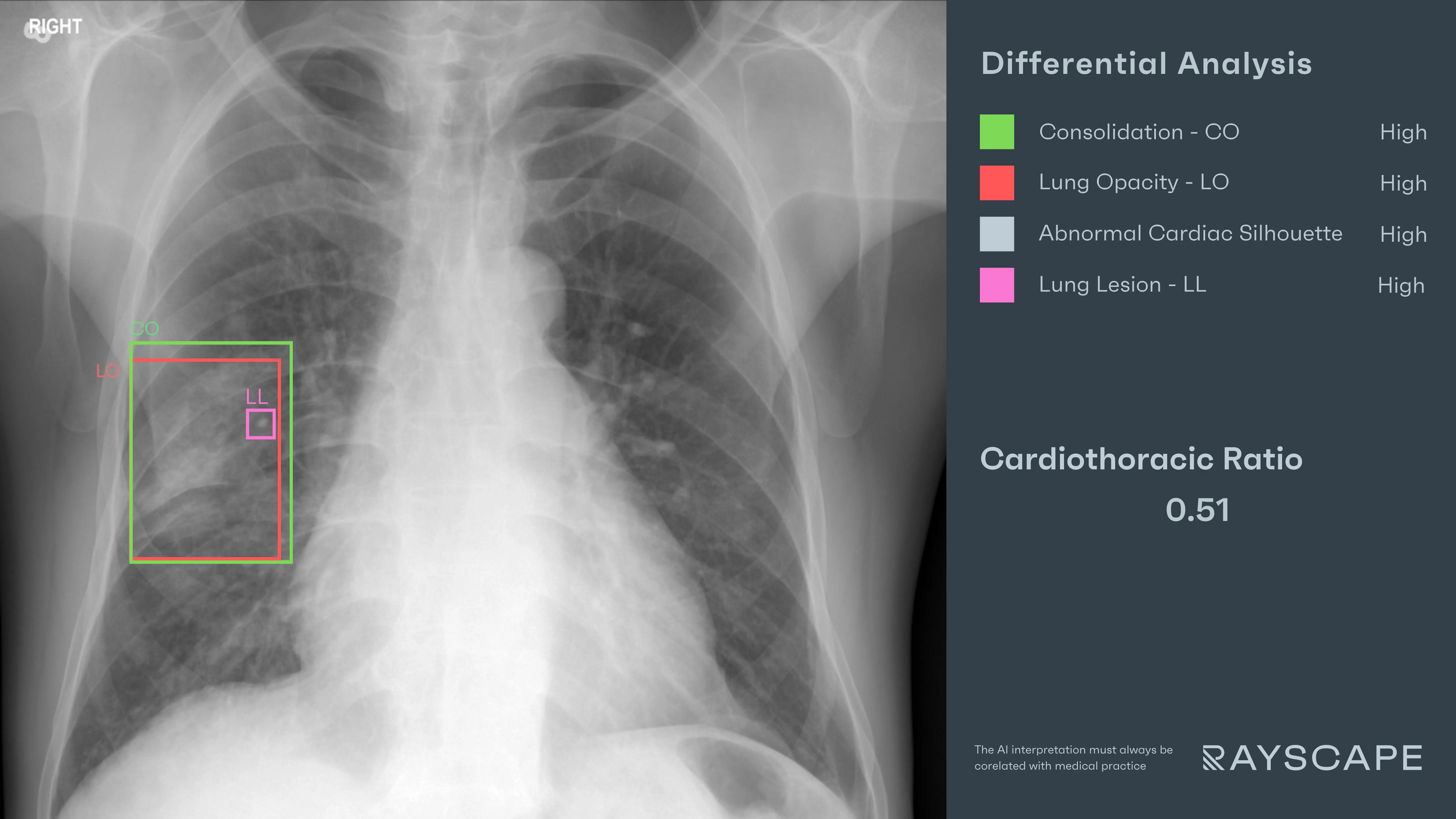
Rayscape ran its analysis in parallel, as it always does—quietly. Then three highlights emerged:
- "Lung Opacity" – A soft, hazy density in the right middle lobe. The kind of opacity that might disappear with a different angle, or a different day.
- "Consolidation" – A denser portion of the same region, confirming the opacity as structured and segmental—not just artifact.
- "Lung Lesion" – Inside the consolidation, the AI outlined a discrete nodule—small, well-circumscribed, and nested within the hazy field.
The radiologist adjusted the contrast. The lung was indeed opacified. And inside it—a distinct round shadow. Without the overlay, it could have passed as part of the infiltrate. But now, framed by the AI, it asked a more specific question: was this post-infectious, or something else entirely?
Outcome
The report was issued with clear recommendations: “Right middle lobe consolidation with embedded nodule. Suggest follow-up imaging post-treatment and CT to characterize underlying lesion.”
A CT was performed one month later. The consolidation had partially cleared, but the nodule remained—7mm, poor-defined, non-calcified. It was entered into a structured lung nodule follow-up protocol.
#OUTPATIENT CASE 3: The lungs were too quiet
Patient: Male, 62 years old
Setting: Outpatient Imaging Center
Clinical Presentation: Referred for fatigue, low-grade fever, and weight loss
Imaging: Chest X-ray
Clinical Context
Sometimes the outpatient center becomes a place where pathology hides in plain sight. The waiting room is quiet. The lighting is soft. The patients answer their names when called.
He had come alone. Filled out the form. “Persistent fatigue. Mild weight loss. Working from home.” No red flags, no acute symptoms. Just another Wednesday morning referral.
AI Interaction
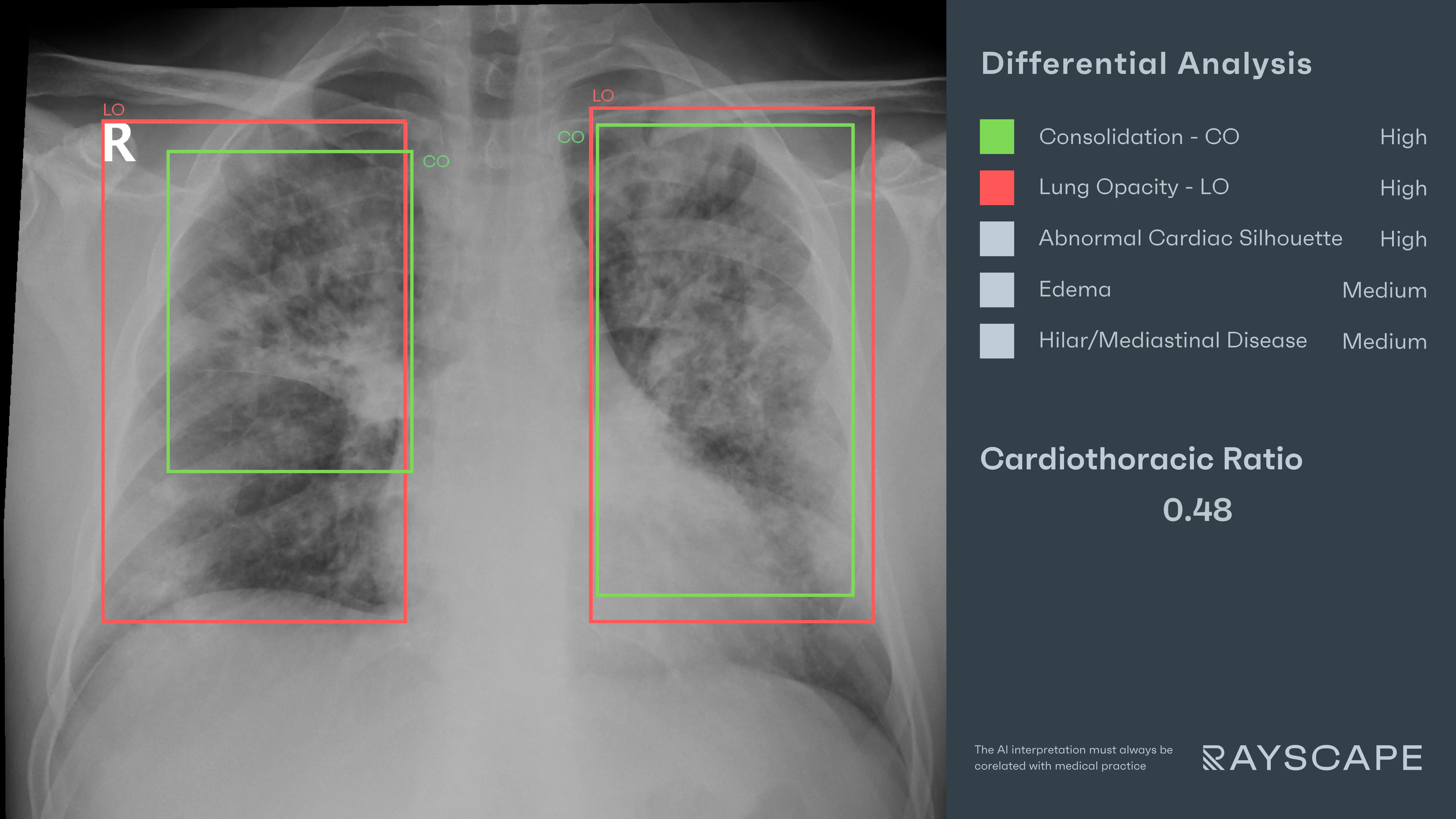
Rayscape stepped in—not to diagnose, but to steady the frame, to sort the noise from the signal.
- "Lung Opacities" – Multiple, scattered across both lungs. Diffuse, patchy, and poorly marginated. Present in every quadrant, with no lobar preference.
- "Consolidation" – A second overlay unified the findings into a larger whole: consolidation involving all lobes, dense and homogenous, with no sparing.
- "Abnormal Cardiac Silhouette" – A soft distortion of the heart’s border, widened at the left base. Not necessarily enlarged—but altered, its contour blending with the adjacent lung shadows.
The radiologist narrowed the window level. The opacities weren’t subtle—they were overwhelming. But somehow the case didn’t scream. It just... settled. The heart looked strange, but not massive. The lungs looked filled, but not specific.
It wasn’t just about what AI detected. It was about visual translation—about giving the radiologist the language to describe it.
Outcome
The report stated: “Diffuse, bilateral consolidations involving all lobes with abnormal cardiac silhouette. Findings concerning for alveolar filling pattern, possibly infectious or interstitial. Recommend immediate CT and clinical reassessment.”
The patient was contacted the same afternoon. A CT scan showed widespread alveolar consolidation with air bronchograms, suggestive of severe pneumocystis pneumonia in an immunocompromised host. Lab tests confirmed previously undiagnosed HIV with low CD4 count.
He was admitted the next morning. Quietly—but just in time.
Discussions
The outpatient center doesn’t move fast—but it moves constantly. No codes, no crash carts—just a quiet stream of patients and films, most of them normal, or close enough to seem that way.
Radiologists here don’t battle chaos. They battle repetition. After enough normal scans, even the abnormal can begin to blend in. Shadows look familiar. Findings feel optional. Attention stretches thin—not from urgency, but from volume and assumption.
In these cases, the AI didn’t diagnose. It didn’t escalate. It framed the quiet signals—subtle consolidations, concealed nodules, displaced silhouettes—before they dissolved into routine.
That’s the role of AI in outpatient care: not to rush the read, but to sharpen attention when everything seems calm. To protect precision from the dulling effect of too many near-normals.
It’s not about speeding up. It’s about not looking away
Built to fit your workflow
For IT teams and PACS administrators, implementation is often a key concern. Rayscape is designed to fit into your existing systems:
- Compatibility with major PACS platforms and cloud environments
- Vendor agnostic: trained to work on every DICOM image, no matter the vendor of the machine or the protocol
- Flexible deployment: on-premise, hybrid, or cloud-based
- Various outputs: SC, GSPS, SR
- Fast onboarding and secure data management protocols (HIPAA / GDPR compliant)
- Available on all major marketplaces (Deepc, Blackford, Carpl) or through direct sale
- We work closely with your radiology and IT departments to ensure seamless rollout with minimal disruption.
Let’s Talk
Your team makes life-saving decisions every day. Rayscape is here to support you in those critical moments—bringing clarity, speed, and confidence to outpatient imaging.
👉 Contact us and learn how we can empower your radiology team—when it matters most.

Discover how Rayscape works
integrate Rayscape AI into
your hospital's infrastructure
and improve both your
daily workflow and patients' experience.
