

AI for Tuberculosis Detection on Chest X-rays
Tuberculosis doesn’t knock loudly. It lingers. It hides. It mimics. And in many regions, X-ray remains the most widely available tool for first-line TB detection—being it ER settings, mobile units, rural outreach programs, and population-level screening caravans.
Here, the challenge isn’t urgency—it’s subtlety. Most patients don’t arrive in crisis. They’re referred for screening, contact tracing, or pre-admission workup. They sit quietly in waiting rooms, their symptoms vague or absent. And radiologists must decide, in seconds, what might become a public health risk tomorrow.
In this setting, the difference between “probably nothing” and “suspicious” can shape isolation decisions, contact protocols, and follow-up care.
Next, you’ll find clinical cases from TB screening programs inside hospital systems—where Rayscape’s AI helped surface key radiological signs of early or atypical pulmonary tuberculosis. Sometimes classic. Sometimes quiet. Always easy to miss without the right frame.
What is Rayscape?
Rayscape is an AI platform trained on millions of medical images. It supports radiologists in detecting a broad spectrum of chest pathologies—including findings often associated with tuberculosis, such as apical opacities, fibrocavitary lesions, nodules, pleural thickening, and volume asymmetry.
In TB screening workflows, whether centralized in hospitals or extended into mobile units, Rayscape acts as a real-time visual assistant—helping radiologists flag abnormalities even when the clinical information is incomplete and image quality isn’t ideal.
It doesn’t diagnose TB. It reinforces attention. It brings subtle, structural changes into sharper focus—before they fade into the noise of another routine film.
The realities of TB screening
Tuberculosis detection often walks a fine line between routine workflow and public health responsibility. These scans aren’t read in trauma bays or isolation wards. They come through quietly—requested for pre-admission, pre-employment, or contact tracing.
And yet, every one of these images carries weight. Because missing TB isn’t just about one patient—it’s about everyone they’ve encountered. Still, the radiologist is faced with familiar obstacles:
- Asymptomatic patients, often with vague or no clinical information
- Quick reads of high-volume chest X-rays, many of which are normal or unrelated
- Findings that are subtle, old, or masked by overlapping anatomy or low-quality images
Here’s how Rayscape makes a difference
- Support radiologists with instant AI-generated highlights that draw attention to overlooked or evolving findings
- Flags early or atypical TB-related findings like apical changes, fibrotic bands, and cavitations
- Surfaces small nodules or streaks that may otherwise be dismissed
- Adds structure and consistency to screening reads, especially in batch workflows
With Rayscape, hospital radiologists gain a second set of eyes—not to replace expertise, but to preserve it, when subtlety meets volume.
Proven clinical value
In the following, you’ll find real-world cases from TB screening programs—where Rayscape supported the detection of pulmonary tuberculosis findings at stages that might otherwise have been missed, delayed, or dismissed.
These examples highlight how AI can assist radiologists in:
- Flagging subtle or atypical signs of early TB, such as small apical opacities, fibrotic streaks, or early cavitations
- Revealing findings hidden behind anatomical structures, using bone suppression to unmask lesions obscured by ribs or the spine
- Maintaining diagnostic vigilance across high-volume reads, even when patient context is minimal and the findings are faint
Each case is anonymized and presented with AI overlays as they appeared in practice. These are not hypothetical scenarios—they are taken from daily workflows, under real clinical pressure.
#TB CASE 1: The weight in the apex
Patient: Male, 55 years old
Setting: Small City Hospital
Clinical Presentation: Persistent fatigue, dry cough, recently arrived migrant with no previous imaging
Imaging: Chest X-ray
Clinical Context
There was no history attached to the request—just a vague phrase typed in all caps: “PRE-ADMISSION CHECK / COUGH.” The patient had filled out his form at reception with the help of a translator. He didn’t speak much. He waited quietly. Thin, upright, tired.
And the X-ray came through just as quietly. Another film in a queue of twenty-five. The kind of day where everything blurs slightly around the edges—where the eyes grow used to normal and the subtle stops being suspicious.
But this one felt different.
The radiologist, six hours into a shift without pause, leaned into the screen. The lungs looked brittle. Not white—but worn. The kind of texture that doesn’t scream—but suggests a long history, unspoken.
This is the real challenge of TB screening in the hospital: the patients don’t look sick. The clinical notes are minimal. And the signs are almost always subtle. The risk isn’t that TB hides—it’s that the system forgets to look for it.
AI Interaction
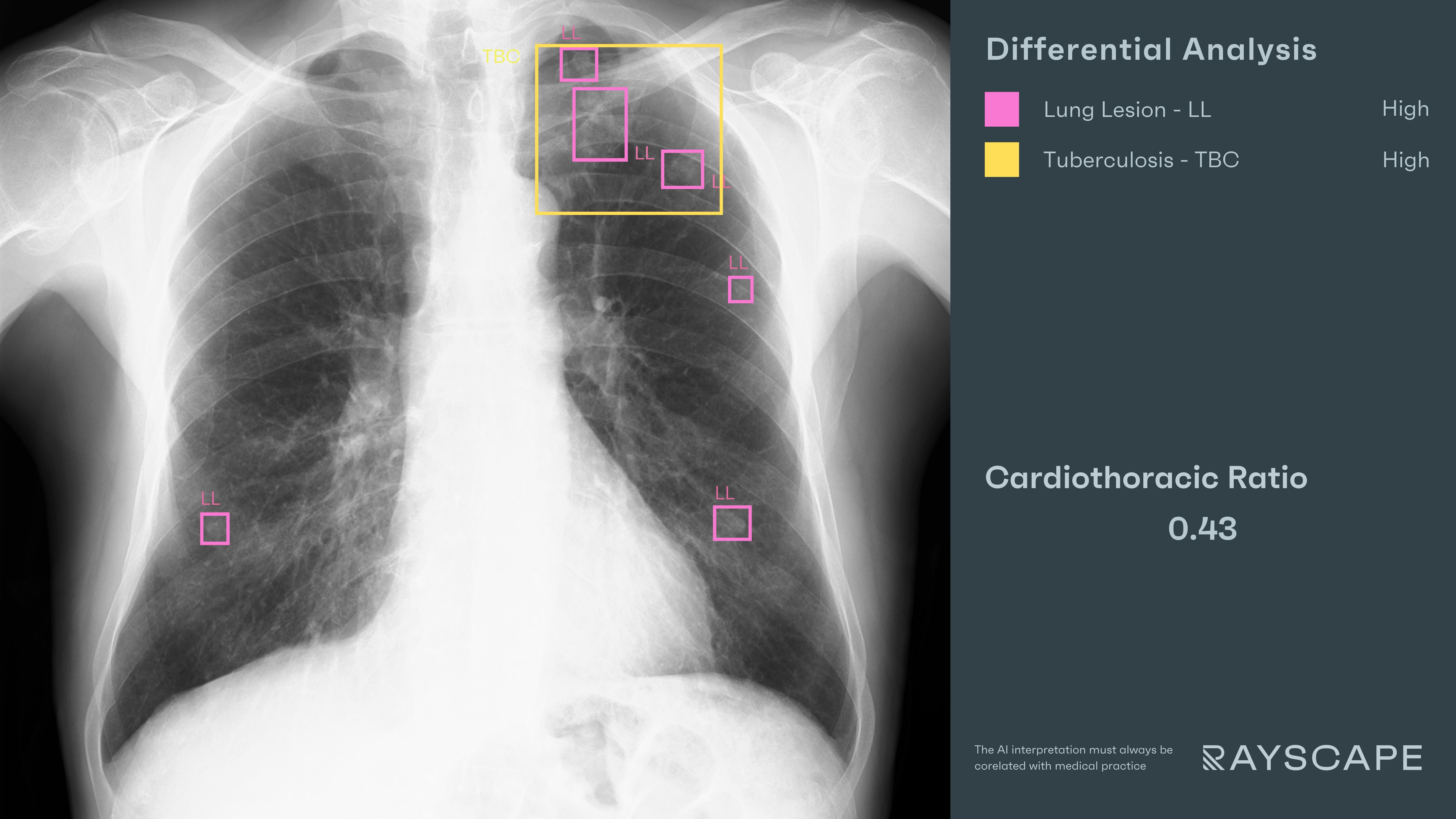
As the chest X-ray loads into the PACS viewer, the Rayscape AI assistant runs its analysis in parallel. Within seconds, its interface overlays distinct bounding boxes:
- "Lung Lesions" – Multiple nodular and reticular densities scattered across both lungs, more densely clustered in the upper zones.
- "Tuberculosis" – A bounding box appeared over the left lung apex, encompassing a complex pattern of fibrotic streaks, architectural distortion, and subpleural reticulation—findings highly suspicious for post-primary or reactivated TB.
No alarms. No urgency. Just structure—a pattern made visible, where fatigue might have blurred the margins.
The radiologist sat forward. These weren’t recent infiltrates. They were textured, fibrotic, laced through the apex like strands of memory. The right lung mirrored smaller changes—but the apex on the left had the weight of history.
The AI does not diagnose, it was the radiologist. What it did was to quietly place the findings in the line of sight—structured, consistent, and visually defined.
Outcome
A GeneXpert test was ordered, followed by CT confirmation. The diagnosis: reactivation of pulmonary tuberculosis, with cavitary transformation not yet visible on X-ray. The patient was started on DOT (Directly Observed Therapy) that same week.
He hadn’t come in expecting a diagnosis. But he received one. Quietly. Early. And just in time.
#TB CASE 2: The fracture wasn’t the whole story
Patient: Male, 48 years old
Setting: Emergency Room
Clinical Presentation: Chest pain post-fall, right-sided tenderness, no fever, stable vitals
Imaging: Portable Chest X-ray
Clinical Context
He came in through the ER—like so many do. Fell on the stairs. Landed awkwardly on his right side. Complained of pain when breathing, but no shortness of breath. The triage note read simply: “Rule out rib fracture.”
And that’s where the mind naturally went. The pain was real. The mechanism was clear. A quick portable X-ray, standard protocol, expected outcome. But ER imaging doesn’t always show what it was asked to find—it sometimes shows what no one thought to ask.
The radiologist was reading from the ER pool. A hundred scans to go. The morning blur of fractures, pneumos, misplaced lines. It would’ve been easy to stop at the obvious. But something else was sitting in the margins—less sharp, less expected.
AI Interaction
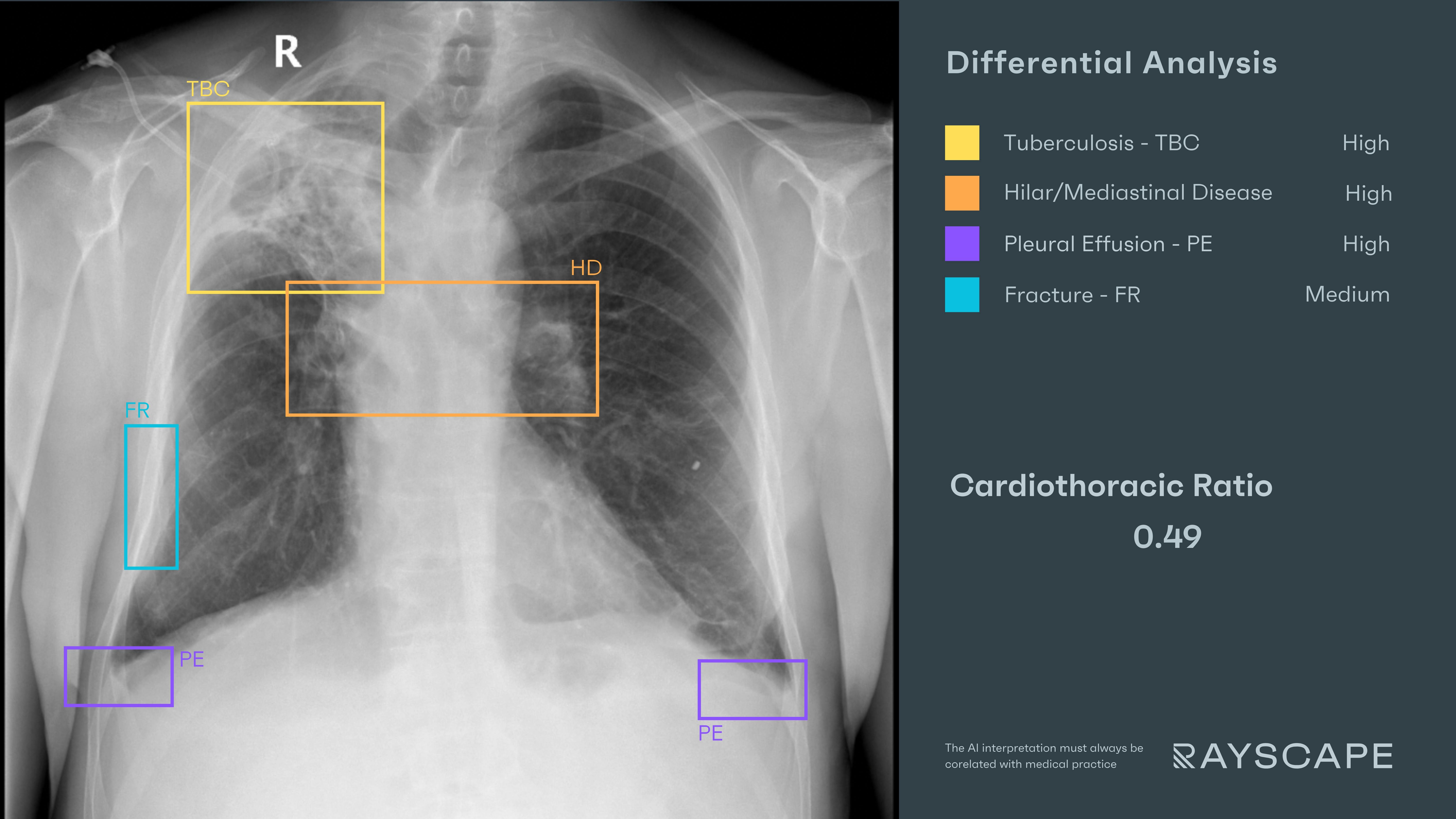
As the image loaded, Rayscape’s AI moved ahead—quietly reading in parallel, not to conclude, but to frame.
- "Rib Fracture" – A faint but clean break. The image was slightly rotated, but the fracture line was undeniable.
- "Pleural Effusion" – Bilateral, small, with more volume on the right, suggesting post-traumatic or reactive fluid.
And then came the unexpected:
- "Tuberculosis" – A structured overlay at the right lung apex, framing a constellation of fibrotic changes, reticulations, and subtle scarring—hallmarks of chronic pulmonary TB.
- "Hilar/Mediastinal Disease" – Another box at the right hilum, capturing soft tissue fullness suggestive of lymphadenopathy or active disease extension.
The radiologist froze. This wasn’t just trauma. This was a lung that had been marked long before the fall. The fracture was acute, yes. But the upper lobe told another story—a story that had been there for years, maybe decades, just waiting for someone to stop and look up from the ribs.
Outcome
The report was updated promptly. The patient was seen the next day by a pulmonologist. GeneXpert was positive. A CT confirmed extensive right apical TB with hilar lymphadenopathy. The fall brought him in—but Rayscape’s AI found more than bone.
And so did the radiologist.
#TB CASE 3: Mirrored, not mild
Patient: Female, 60 years old
Setting: Hospital-based TB Screening Program
Clinical Presentation: Fatigue, intermittent cough, no prior imaging, lives in a high-incidence area
Imaging: Chest X-ray
Clinical Context
There’s a peculiar danger in symmetry—it tricks the brain into complacency. If something looks the same on both sides, it must be something else.
She came in for a routine TB screening. No fever. No weight loss. Just a long cough and a primary care referral. The radiologist had already read three pre-ops and a dozen normal chests that morning. This one looked… patterned. Balanced. But not clear.
AI Interaction
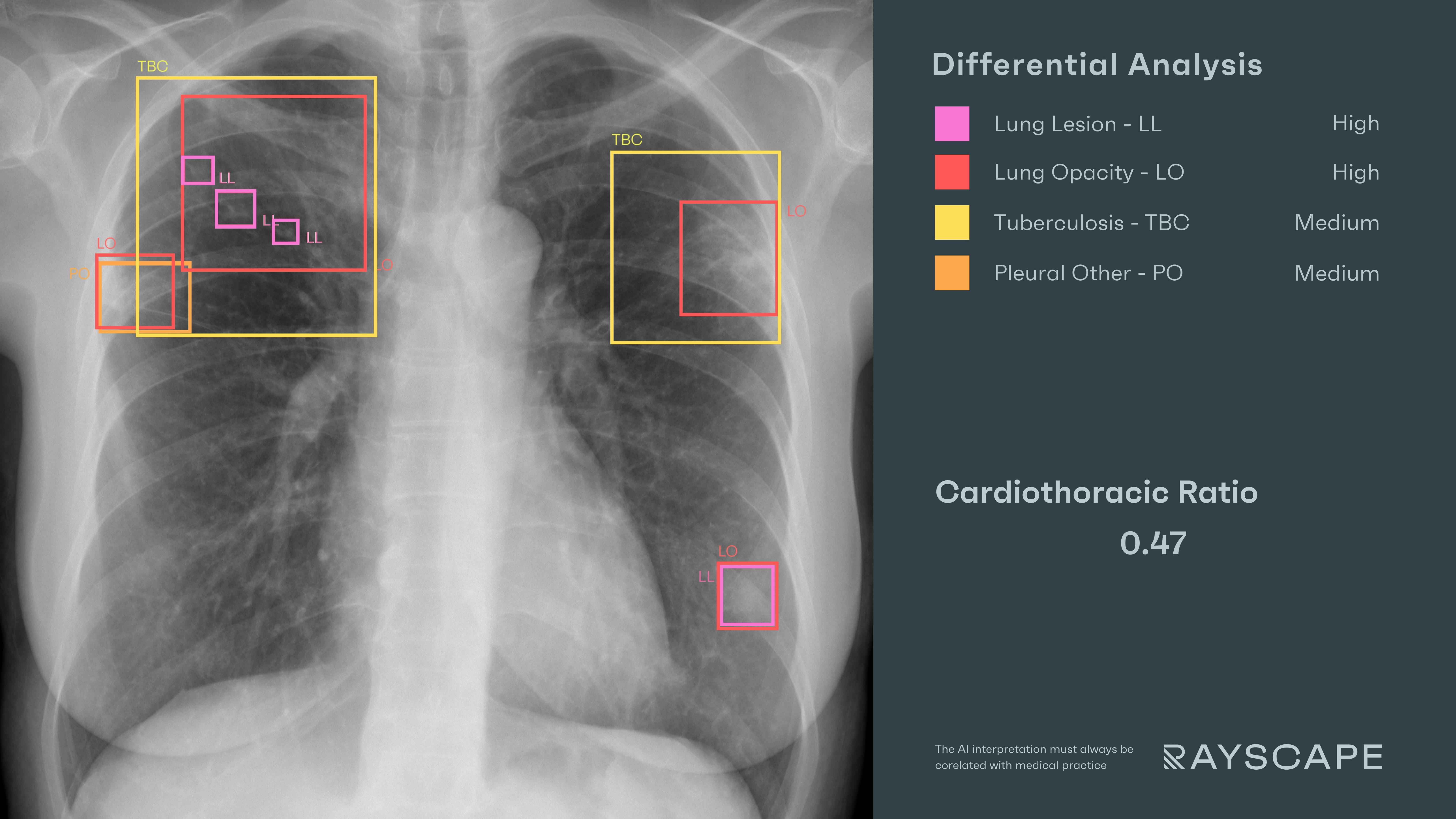
Rayscape processed the image with its usual quiet precision and began surfacing what the symmetry had nearly masked:
- "Lung Lesions" – Multiple bilateral nodular lesions, irregular in shape and distributed across both upper and lower-lung fields.
- "Lung Opacities" – Faint reticulations and parenchymal haziness
- "Tuberculosis" – Structured overlays appeared in upper zones—left and right—capturing areas of fibrotic change, subpleural scarring, and architectural distortion consistent with chronic TB.
It wasn’t just one nodule. It wasn’t just one lung. It was a textbook TB lung. The AI suggested a pattern—and framed it before fatigue could excuse it away.
Outcome
The report flagged: “Bilateral upper-lobe nodular lesions with apical fibrosis and reticulations. Findings consistent with chronic pulmonary tuberculosis.” The patient tested positive. CT confirmed cavitations and more extensive disease than seen on X-ray alone. Treatment was initiated the same week.
Discussions
Tuberculosis doesn’t rush—but it leaves a mark. And the radiologist’s challenge is not just to spot it, but to recognize it when nothing else insists on being noticed.
These are not trauma films. There are no bleeds, no fractures, no codes. These are lungs that carry a quieter burden—etched into apices, hidden behind bones, mirrored across both fields in patterns easy to misread as age, artifact, or scar.
But what’s easy to explain is not always safe to ignore.
In these cases, Rayscape helped frame what fatigue might flatten, and what busy workflows might wave past. It slowed the scroll. It refocused the frame.
This is where AI finds its place in TB screening: not in shouting, but in asking the right questions with quiet precision. In offering radiologists a pause—a second glance at findings that, left alone, might become someone else’s burden to carry.
The future of TB detection won’t come from louder machines. It will come from sharper attention, consistently applied, case after case—even when the lungs look symmetrical, the histories are thin, and the findings feel familiar.
Built to fit your workflow
For IT teams and PACS administrators, implementation is often a key concern. Rayscape is designed to fit into your existing systems:
- Compatibility with major PACS platforms and cloud environments
- Vendor agnostic: trained to work on every DICOM image, no matter the vendor of the machine or the protocol
- Flexible deployment: on-premise, hybrid, or cloud-based
- Various outputs: SC, GSPS, SR
- Fast onboarding and secure data management protocols (HIPAA / GDPR compliant)
- Available on all major marketplaces (Deepc, Blackford, Carpl) or through direct sale
- We work closely with your radiology and IT departments to ensure seamless rollout with minimal disruption.
Let’s Talk
Your team makes life-saving decisions every day. Rayscape is here to support you in those critical moments—bringing clarity, speed, and confidence to emergency imaging.
👉 Contact us and learn how we can empower your radiology team—when it matters most.
Discover how Rayscape works
integrate Rayscape AI into
your hospital's infrastructure
and improve both your
daily workflow and patients' experience.
