

How radiologists use AI in lung cancer screening
The rising volume of low-dose CT scans in lung cancer screening is creating a new challenge: one where subtle findings must be tracked, compared, and scored with precision across time.
While CT imaging continues to improve, the challenge is no longer just detection—it’s standardization, recall, and longitudinal clarity. This is especially true in high-throughput screening programs, where missed evolution can delay care, and false escalation can cause unnecessary anxiety.
Next, we invite you to explore real cases where Rayscape’s AI didn’t replace the radiologist—but acted as a perceptive observer: framing what’s subtle, structuring what’s scattered, and helping decisions land with greater clarity.
Seeing earlier, acting smarter in Lung Cancer Screening
In lung cancer screening programs, precision matters more than urgency. Radiologists must interpret a growing number of low-dose CT scans, often with limited clinical context—while navigating a forest of nodules, most of which won’t require action.
Whether to reassess in 12 months or move to biopsy. And the reality is that:
- Nodules are often small, stable, or obscured by overlapping anatomy
- Comparisons across time can be inconsistent, especially when protocols differ
- Even experienced radiologists may struggle with volume tracking or scoring edge cases
Rayscape supports this process by:
- Highlighting nodules automatically, with segmentation and volume measurement
- Calculating Lung-RADS categories based on nodule size, type, and evolution
- Surfacing changes between timepoints with structured overlays
- Ensuring visual consistency across baseline and follow-up scans—without disrupting workflow
With Rayscape, radiologists can focus on clinical judgment and context—while we help ensure that what matters doesn’t slip through the cracks.
Learn more below from real clinical cases from lung cancer screening programs across multiple institutions—each one illustrating how AI has supported radiologists in detecting, measuring, and tracking findings such as:
- Pulmonary nodules
- Nodule growth across time points
- Lung-RADS category changes
- ...and more
Each case is anonymized and presented with AI overlays as they appeared in practice. These are not hypothetical scenarios—they reflect real workflows, under the quiet but critical pressure of screening reads.
#SCREENING CASE 1: A shadow that grew in silence
Patient: Male, 64 years old
Setting: Lung Cancer Screening Follow-up Program
Clinical Presentation: Asymptomatic
Imaging: Baseline CT + follow-up CT after 12 months
Clinical Context
The thing about screening is that nothing ever seems urgent. No red flags. No crisis. Just list after list of low-dose CTs—repetitive, routine, and painfully easy to forget.
He was 64. No smoking in the past ten years but history of asbestos work exposure. The scan looked like so many others: clean lung fields, a handful of nodules.
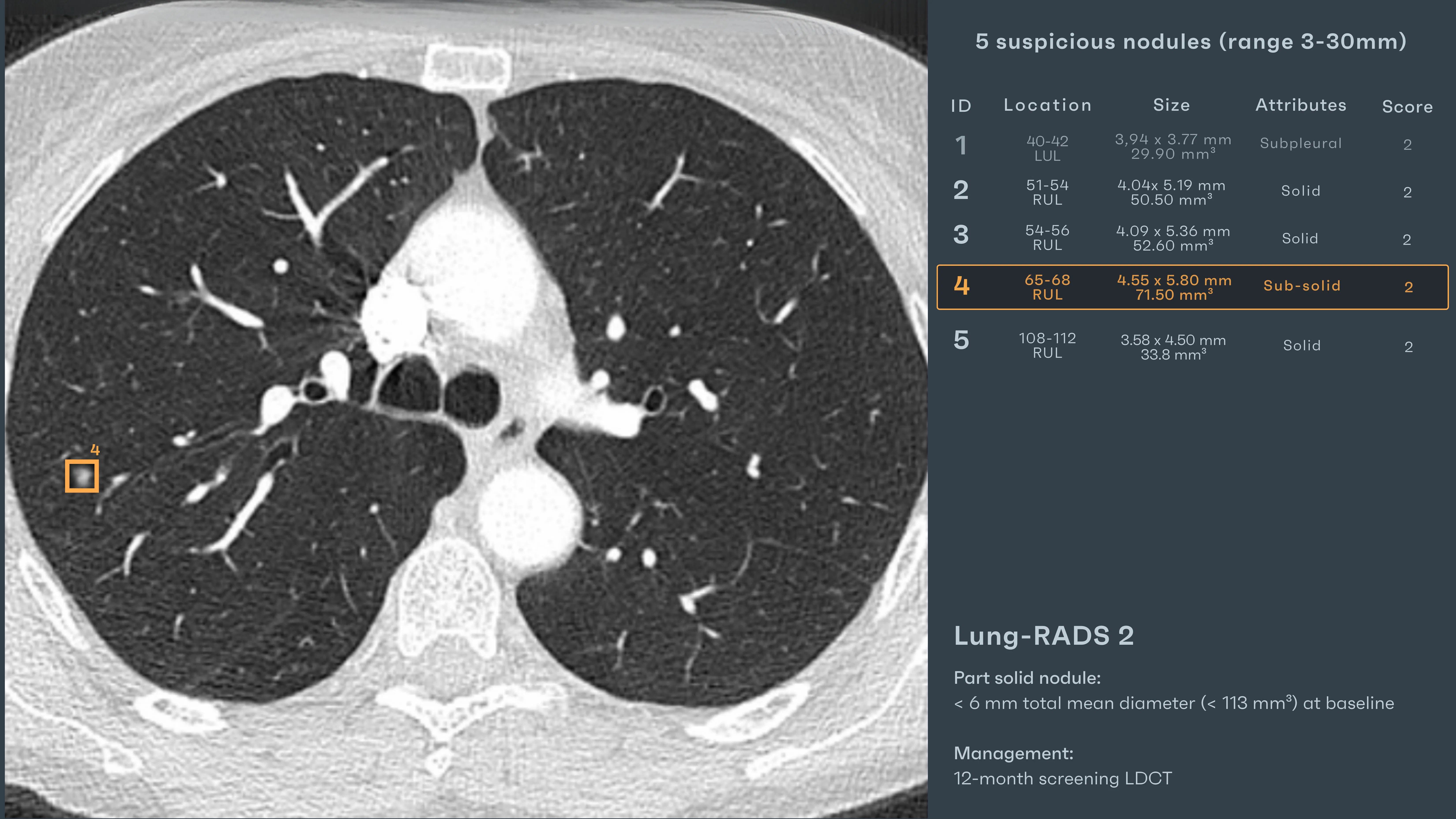
Four were round and small. But one—subsolid, near the pleura—lingered. 4.5 by 5.8 mm. Smooth margins. No obvious risk signs. Rayscape AI assigned Lung-RADS 2, with a 12-month recall.
A year later, the follow-up appeared at the bottom of the list. Nothing flagged it. No red dot. No comment in the referral. Just another case, waiting. And this is where it happens: not in the moment of crisis, but in the space between.
Between last year’s almost-nothing and this year’s maybe-something. Between what was recorded, and what was remembered.
AI Interaction
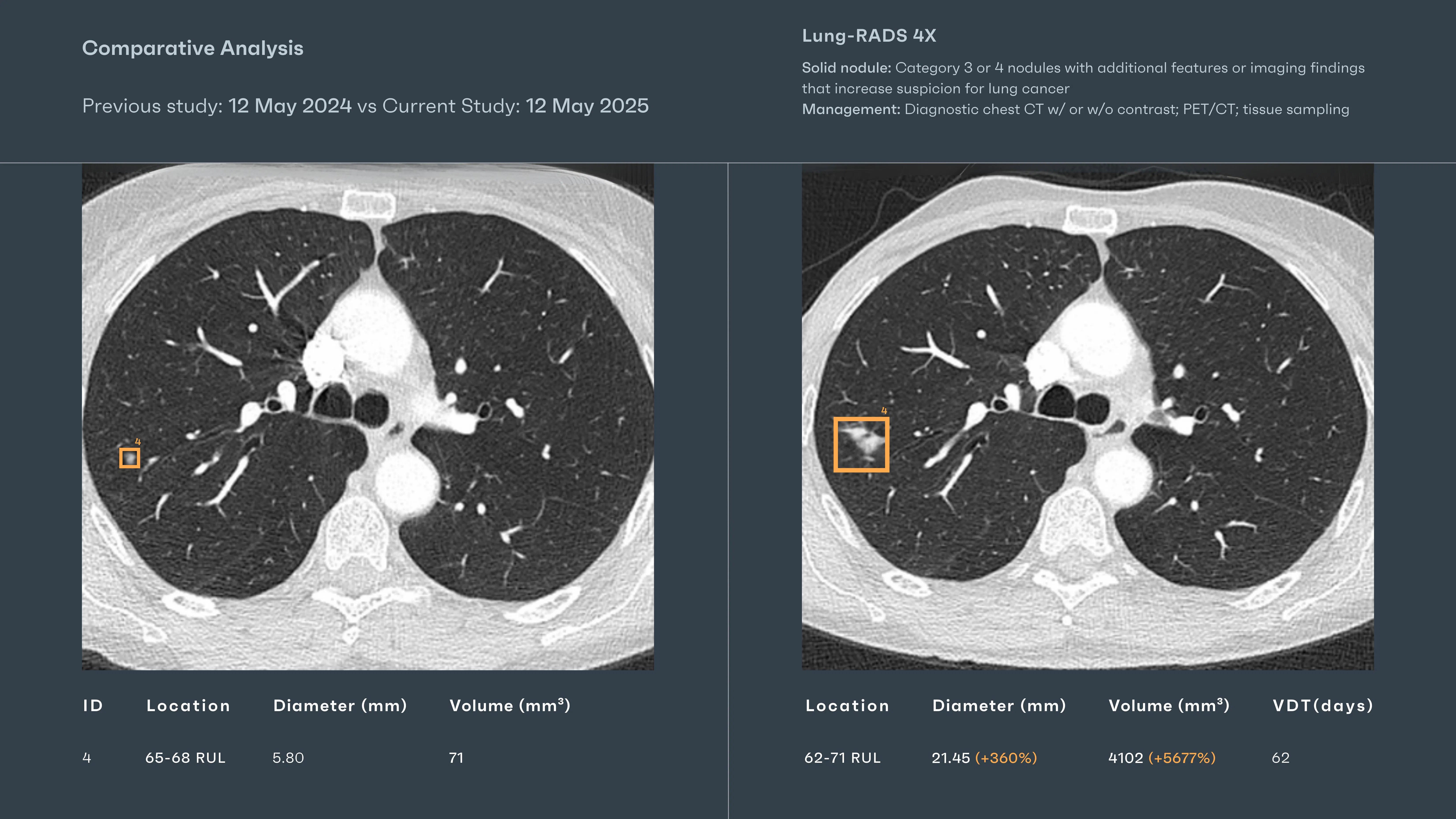
As both CT scans loaded, Rayscape began processing. Within seconds, it placed visual overlays directly on the comparison view:
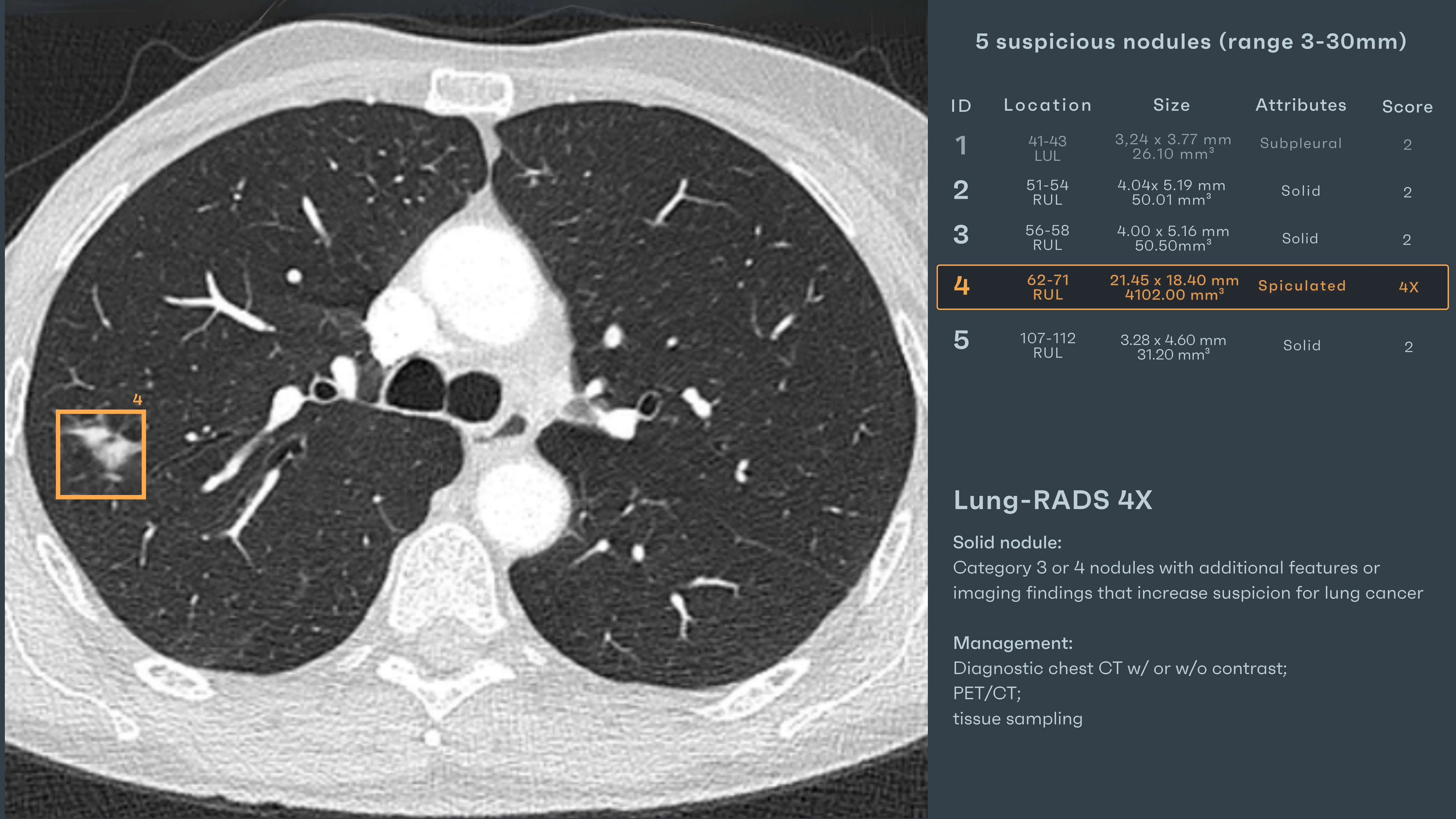
- The nodule in the right upper lobe, now 21.45 × 18.40 mm, with a measured volume of 4102 mm³, up from 71.5 mm³ a year earlier
- New spiculated margins detected
- System-generated Lung-RADS 4X score, based on size, morphology, and change over time
The AI didn’t suggest urgency. It didn’t diagnose malignancy. It presented structure: growth, risk, category—visually clear, quantitatively precise, algorithmically grounded. The radiologist reviewed the overlay, scrolled through axial cuts, adjusted the window. The findings held.
#SCREENING CASE 2: A simple nodule
Patient: Female, 62 years old
Setting: Lung Cancer Screening Program
Clinical Presentation: Asymptomatic, Smoking history
Imaging: Baseline CT
Clinical Context
There’s a peculiar tension in lung screening: the fear of missing something, and the fatigue of seeing nothing. Most nodules stay quiet. Most follow-ups are routine. But the radiologist doesn’t get to know that until after the second scan.
She was 72, referred for baseline CT as part of a enrollment into the screening program. No symptoms. Just smoking history. Another scan at the end of a long day—another moment of potential tucked inside clinical routine.
The image looked clean. The lungs were mostly clear. But the thing about screening
isn’t what you expect—it’s what you might miss by assuming.
AI Interaction
As the CT loaded, Rayscape’s AI processed the image in parallel, presenting overlays
directly in the radiologist’s line of sight:
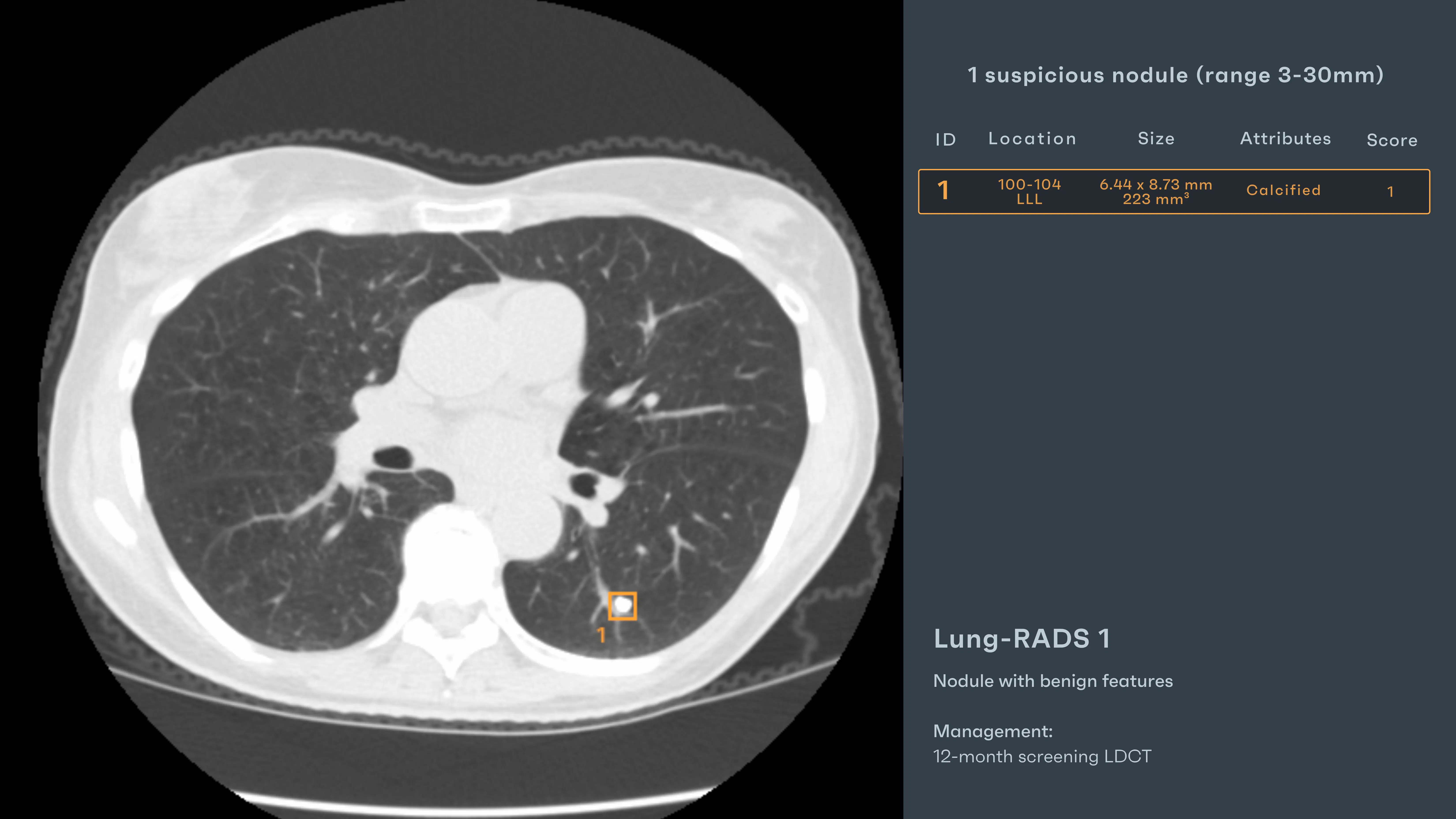
- "Calcified Nodule" – Left lower lobe, 6.44 × 8.73 mm, 223 mm³, solid, dense, central calcification pattern
- System-generated Lung-RADS 1 – Based on morphology and measurements
Rayscape outlined a narrative, not as an alarm, but as a quiet confirmation: a calcified contour, smooth margins, dense core—hallmarks not of urgency, but of time. The radiologist paused, reviewed axial and coronal views, adjusted the windowing—a benign presence, steady and self-contained.
Outcome
The report was finalized without escalation. The AI-generated Lung-RADS 1 score was accepted, and the recommendation noted: annual low-dose CT, per protocol. At the next evaluation, the nodule remained unchanged—no growth, no transformation. Just the same story, retold with quiet certainty.
#SCREENING CASE 3: The weight of a maybe
Patient: Male, 66 years old
Setting: Lung Cancer Screening Program
Clinical Presentation: Former smoker, asymptomatic
Imaging: Baseline CT only
He came in on schedule. Former smoker, no symptoms, just a routine CT booked months in advance. It was one of those scans that arrived between two follow-ups and a post-COVID fibrosis read—quiet, untitled, expected. But this one didn’t slide easily into the rhythm of routine.
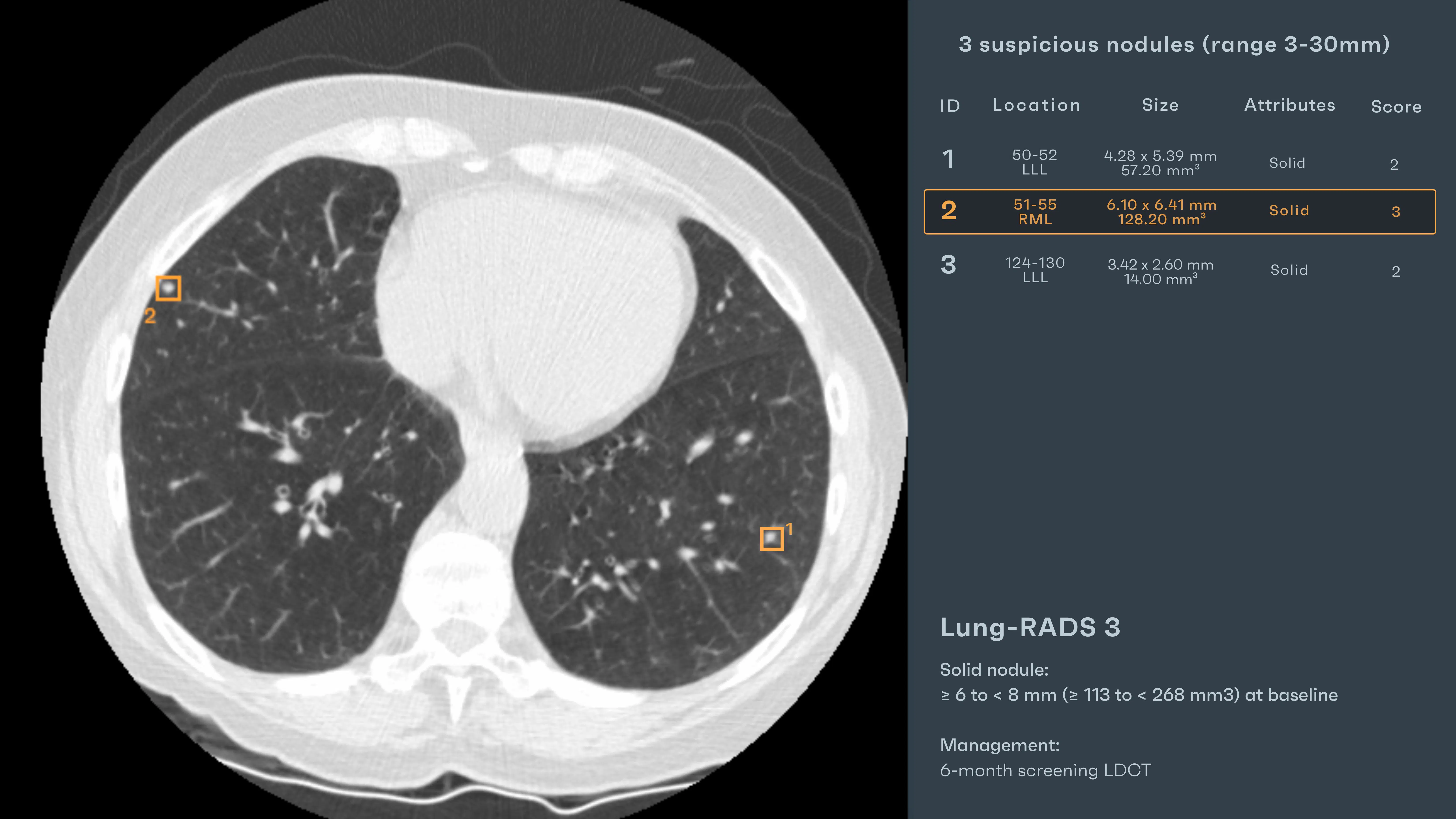
Three nodules.
One in the LLL, small and well-defined, a 2nd one in RML and a 3rd in LLL. They could have been easily missed. But it was the second one—, solid and distinct—that held the screen just a little longer.
No spiculation. No fat. But enough presence to feel wrong in its stillness. This is where the pressure builds—not in the volume of scans, but in the quiet ones that leave just enough doubt. Simple measurements where a mouse sensitivity can slip and show a smaller dimension from the radiologist point of view. A change that could impact the future of the patient.
AI Interaction
Rayscape processed the scan in parallel. Overlays appeared with structured clarity:
- "Solid Nodule #1" – Left lower lobe, 5.3 mm, rounded, smooth
- "Solid Nodule #2" – Right middle lobe, 6.10 × 6.41 mm, 128.20 mm³, well-circumscribed
- "Solid Nodule #3" – Second LLL nodule, sub-centimeter size
The AI System-generated Lung-RADS 3 – Based on size and morphology.
There was no call to act immediately. No overstep. The system placed structure beside instinct. The nodule wasn’t large, but it was precise.
Outcome
The report noted: “Three solid nodules, most prominent in RML, measuring 6.10 × 6.41 mm. AI-generated Lung-RADS 3. Recommend repeat LDCT in 6 months.”
At follow-up, the right middle lobe nodule had grown. Edges sharpened. Volume increased. PET/CT confirmed uptake. Biopsy followed. The diagnosis: adenocarcinoma.
The system had framed it early. The radiologist had seen. And the patient, caught in that space between maybe and malignancy, got something a chance to act.
Discussions
Radiologists aren’t tireless or immune to overload. Behind every lung CT report is a reader juggling timelines, growth patterns, and nuanced findings—while the next scan waits in the queue. The pathology wasn’t hidden. It lived in ambiguity: a borderline nodule, a subtle ground-glass opacity, a shift just below the threshold of alarm.
The AI didn’t “catch” what the radiologist missed—it offered clarity before the fog could form. It positioned the findings within the frame, allowing the mind to move faster, the pattern to resolve more sharply. And this is the quiet revolution: not replacement, but reinforcement. Not autonomy, but augmentation. Rayscape acted not as a second opinion, but as a second set of steady hands in a moment where one pair may not be enough.
The future of lung CT is collaborative. Where human intuition meets algorithmic precision. Where a bounding box becomes not a conclusion, but a question, well-posed, in just the right place.
Built to fit your workflow
For IT teams and PACS administrators, implementation is often a key concern. Rayscape is designed to fit into your existing systems:
- Compatibility with major PACS platforms and cloud environments
- Vendor agnostic: trained to work on every DICOM image, no matter the vendor of the machine or the protocol
- Flexible deployment: on-premise, hybrid, or cloud-based
- Various outputs: SC, GSPS, SR
- Fast onboarding and secure data management protocols (HIPAA / GDPR compliant)
- Available on all major marketplaces (Deepc, Blackford, Carpl) or through direct sale
- We work closely with your radiology and IT departments to ensure seamless rollout with minimal disruption.
Let’s Talk
Your team makes life-saving decisions every day. Rayscape is here to support you in those critical moments—bringing clarity, speed, and confidence to outpatient imaging.
👉 Contact us and learn how we can empower your radiology team—when it matters most.

Discover how Rayscape works
integrate Rayscape AI into
your hospital's infrastructure
and improve both your
daily workflow and patients' experience.
