

Rayscape × Bucharest Emergency University Hospital Collaboration
Lung cancer remains one of the most serious public health challenges in Romania, with late diagnosis continuing to drive poor outcomes. In the absence of a national screening program, many lung cancers are still discovered incidentally, during imaging performed for unrelated clinical reasons. In this reality, radiology becomes a critical frontline, often holding the first opportunity to change a patient’s trajectory.
Emergency hospitals sit at the center of this challenge. They operate under constant pressure, manage high imaging volumes, and serve patients with acute, complex, and oncological conditions around the clock. For radiologists working in these environments, speed and accuracy are equally essential—and increasingly difficult to balance.
Within this context, Rayscape partnered with the Bucharest University Emergency Hospital (SUUB), with the support of AstraZeneca, to explore how artificial intelligence can be integrated into real-world clinical workflows to support earlier detection, consistent evaluation, and longitudinal monitoring of lung disease.
The challenge: imaging under continuous pressure
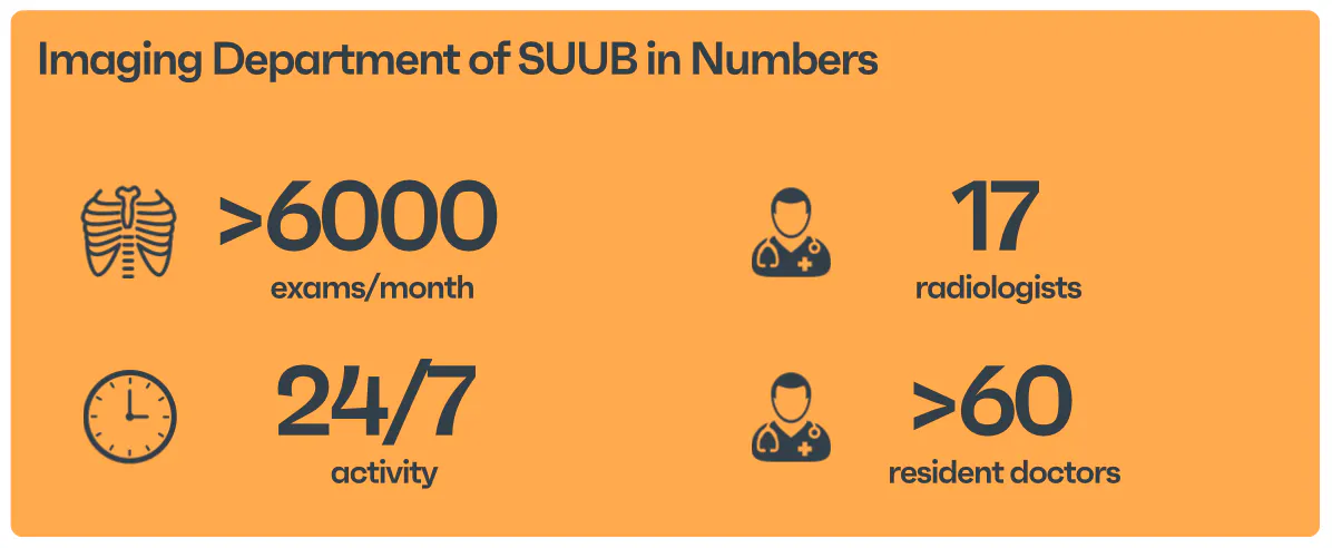
In emergency and university hospital settings, chest X-rays and CT scans are performed at scale, often to answer urgent clinical questions such as trauma, acute respiratory symptoms, or post-operative complications. Radiologists must prioritize life-threatening findings, while subtle pulmonary nodules may fall outside the immediate clinical focus.
Manual interpretation under time pressure, combined with cognitive load and visual fatigue, increases the risk that incidental but clinically significant findings remain unreported or insufficiently followed up. This is particularly relevant in lung cancer, where early-stage disease may present as small, asymptomatic nodules.
As imaging volumes grow and staffing constraints persist, these limitations become structural rather than individual.
The solution: AI embedded directly into clinical workflow
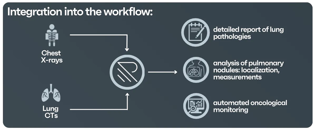
At SUUB, artificial intelligence was integrated directly into the existing PACS infrastructure, without altering acquisition protocols or reporting routines. Rayscape analyzes all chest X-rays and lung CT scans automatically, in the background, and returns results within the same study.
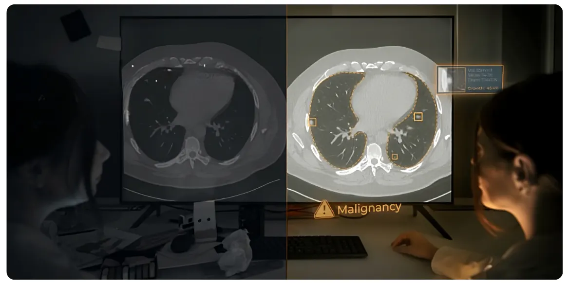
For chest X-rays performed in the Emergency Department and radiology units, AI highlights thoracic abnormalities and incidental pulmonary nodules, supporting radiologists in prioritization and documentation. For lung CT examinations, the system automatically detects and measures nodules, applies Fleischner guideline recommendations, and flags patients requiring structured follow-up.
Importantly, AI also evaluates pulmonary bases visible on abdominal CT scans, an area where incidental findings are commonly overlooked, reducing the risk of diagnostic omission.
From detection to longitudinal monitoring
Beyond single-exam analysis, the collaboration focused on longitudinal assessment. For patients undergoing oncological monitoring or follow-up imaging, AI aligns studies across time, matches corresponding lesions, and calculates objective metrics such as volume, diameter, growth rate, and volume doubling time.
This standardized approach supports consistent evaluation across departments and over time, reducing variability and enabling clearer communication between radiology and oncology teams. Radiologists remain fully in control of interpretation, while benefiting from objective, reproducible data.
Toward measurable clinical impact
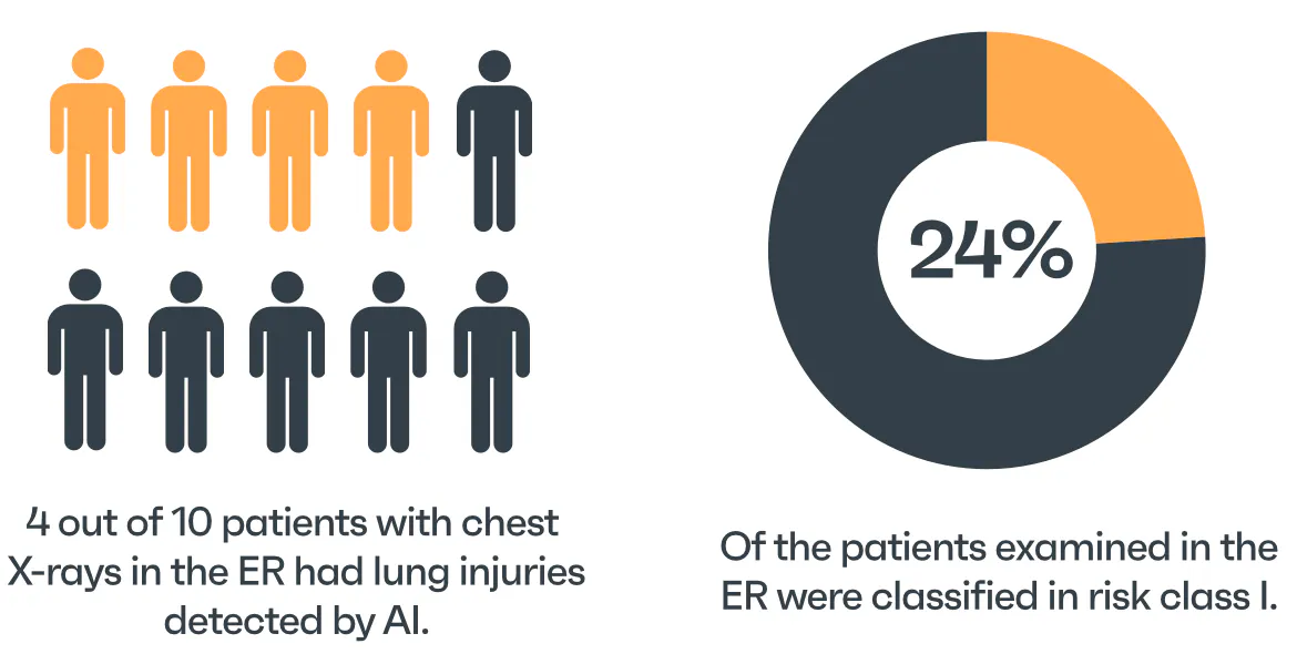
SUUB demonstrates how artificial intelligence can move beyond experimental deployment and become a practical clinical support tool in high-pressure hospital environments. By making incidental findings visible, measurable, and traceable, AI helps transform routine imaging into an opportunity for earlier intervention, particularly in a healthcare system without population-wide lung cancer screening.
Want to explore the full case study and technical details behind this collaboration?


Discover how Rayscape works
integrate Rayscape AI into
your hospital's infrastructure
and improve both your
daily workflow and patients' experience.
