

Best AI Companies for Lung Cancer CT Screening in 2026
Low-dose computed tomography (LDCT) screening has become a critical public-health tool because lung cancer is far more curable when detected early. Interpreting these volumetric scans is labor-intensive and prone to oversight; radiologists must review hundreds of slices and characterize small pulmonary nodules that may indicate malignancy. AI-based imaging solutions are reshaping patient care by improving diagnostic accuracy while also supporting cost control and clinical safety.
With ongoing technological progress and the use of large-scale datasets that refine algorithm performance, diagnostic imaging has entered a new phase of capability in recent years. As AI-supported radiology becomes an integral component of lung cancer screening programs, several commercial AI systems now automate nodule detection, characterization, and follow-up on chest CT while fitting into existing clinical workflows. The following companies represent key radiology vendors active in AI solutions for lung cancer screening in 2026, highlighting the expanding momentum in this domain.
Rayscape – the Radiology AI for Lung CT & Lung‑Cancer Pathway
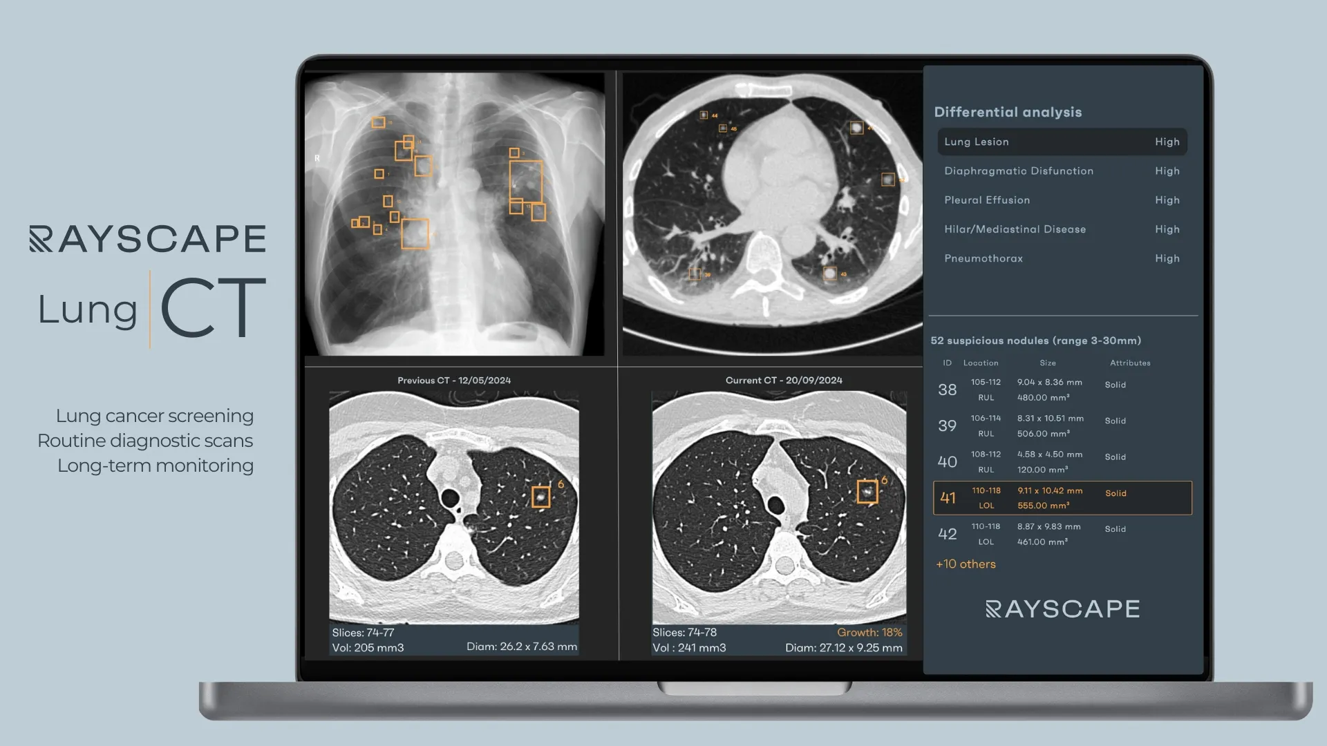
Rayscape built the Lung CT module to streamline the practical workflow of lung-cancer screening and follow-up. The algorithm highlights nodules between 3 and 30 mm, records their lobar locations, and automatically measures their diameters and volumes in three orthogonal planes. The AI developed by the Rayscape team is designed to work across all scanning variations: non-contrast or contrast, normal dose or low-dose (LDCT), and it helps with:
- Detection range and localisation: Besides identifying and providing a list for each suspected nodule’s location, the model marks its lobar location on CT slices, helping radiologists quickly focus on regions of interest.
- Automated measurements and attributes: For every nodule, Lung CT automatically measures diameter and volume in three orthogonal planes and assigns a set of attributes such as texture (calcified, solid, sub-solid, etc.), and margin (such as spiculated) or position (subpleural or not). These measurements enable standardised baseline characterisation.
- Growth analysis and reporting: On prior/subsequent scans, the AI matches previously detected nodules, compares their size and volume, and generates growth reports with guideline-based recommendations aligned with Fleischner, BTS, or Lung-RADS criteria. Reports are customisable and can be integrated both into PACS and local systems, or visualized in the web viewer, with automated population of key nodule descriptors, guideline categories, and follow-up recommendations, reducing reliance on manual transcription or separate workstations and promoting standardized communication.
- Workflow integration: Rayscape emphasises “plug-and-scan” deployment; the AI integrates with existing infrastructure, whether on-premise or cloud-based, and smart reports provide both visual and textual outputs.
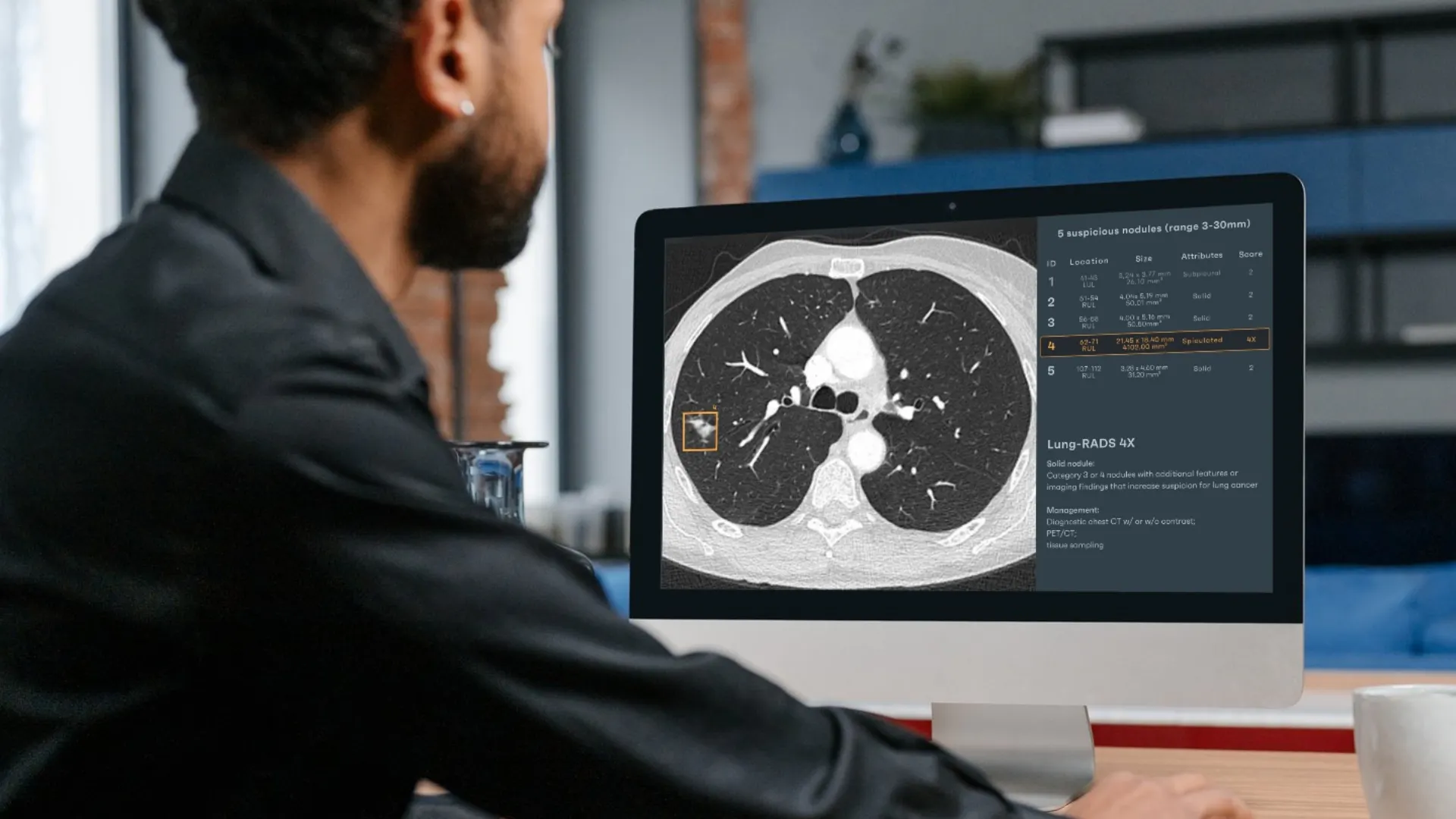
Rayscape’s Lung-Cancer Pathway extends the core CT module into a longitudinal care platform. The suite monitors incidental nodules that may be found even on abdominal CTs by reanalyzing the lung bases and sends automatic follow-up recommendations. It incorporates bone-suppressed chest X-ray analysis to detect nodules that are difficult to see when ribs or clavicles obscure pulmonary tissue, and it unifies findings across X-ray and CT scans. The system calculates volume-doubling time (VDT), standardises report formatting, and aligns follow-up intervals, turning Rayscape from a spot-detection module into a full lung cancer pathway tool for incidental finding management, screening, and oncology tracking and follow-up.
Coreline – AVIEW LCS
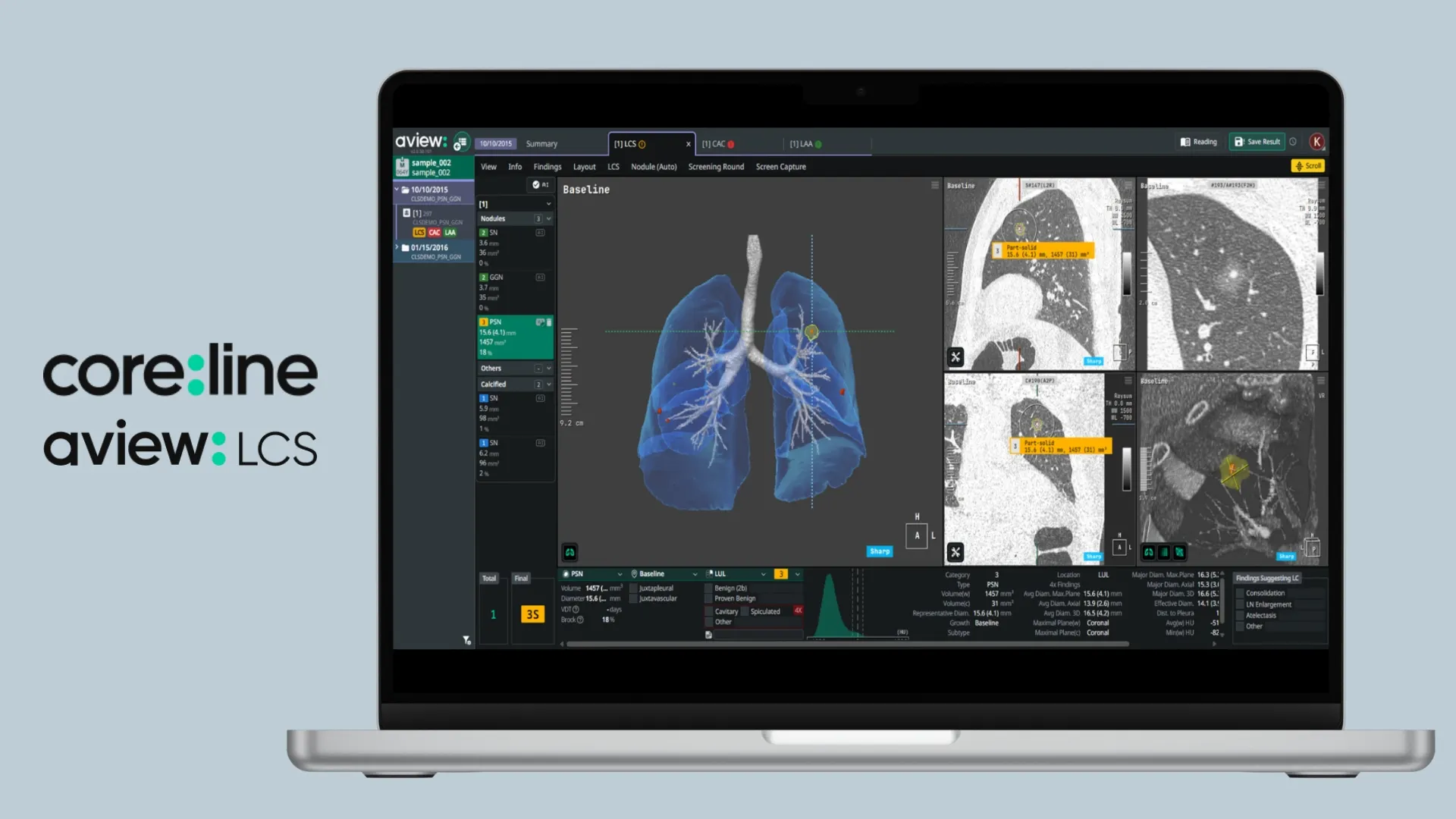
Coreline’s AVIEW LCS is an AI lung CT screening solution designed to support low-dose CT (LDCT) screening workflow through automated detection, characterization, and longitudinal analysis. The system focuses on identifying pulmonary nodules on screening chest CT and structuring their evaluation in line with Lung-RADS–based management pathways.
At the detection and characterization level, the deep-learning engine identifies pulmonary nodules and performs three-dimensional segmentation, enabling volumetric analysis. Nodules are categorized by attenuation profile—solid, part-solid, or non-solid—and the system records key parameters such as volume, diameter, and lesion count. These quantitative outputs form the basis for standardized baseline documentation and risk-oriented categorization.
AVIEW LCS incorporates automated Lung-RADS classification logic aligned with the American College of Radiology framework. Based on nodule size, type, and growth behavior, the system assigns categories that are directly linked to recommended follow-up strategies, embedding guideline logic into the reporting process rather than treating it as a separate manual step.
A dedicated longitudinal module supports follow-up imaging. The software automatically registers current and prior CT examinations, matches corresponding nodules across timepoints, and evaluates interval changes using volumetric growth metrics. This longitudinal tracking is intended to support consistent assessment of stability, progression, or interval development of new nodules within structured screening programs. From a visualization and workflow perspective, AVIEW LCS includes a clinical viewer with 3D lung rendering, allowing users to review segmented nodules within a volumetric anatomical context.
contextflow – ADVANCE Chest CT
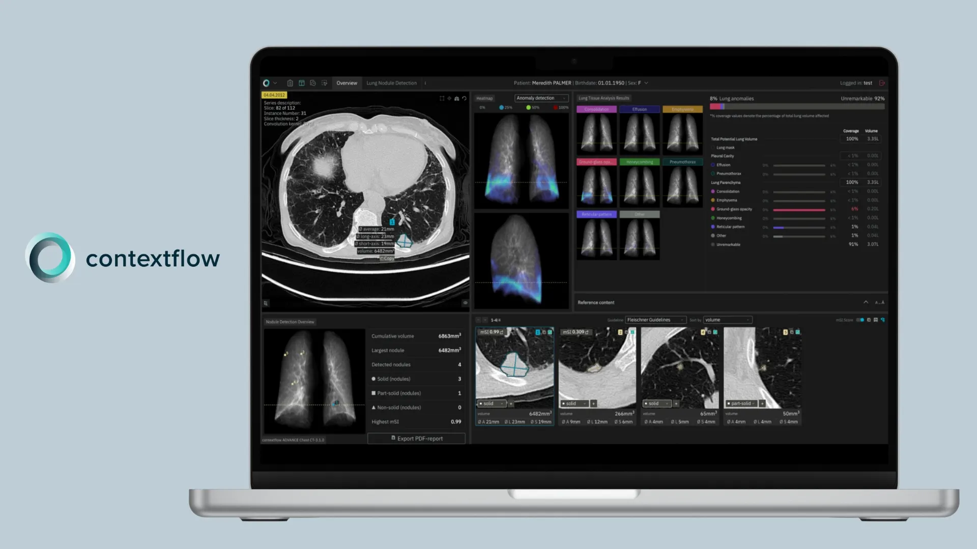
Contextflow is an Austrian startup that created ADVANCE Chest CT, a CAD module that focuses on nodule detection and lung‑tissue quantification. It detects and quantifies nodules between 4 and 30 mm and classifies them as solid, part‑solid, or non‑solid. The system tracks these findings over time and produces a nodule‑tracking report. Beyond nodules, ADVANCE Chest CT segments the lung and quantifies patterns such as consolidation, effusion, emphysema, ground‑glass opacity, honeycombing, pneumothorax, and reticular pattern. These quantitative maps are captured as DICOM secondary captures so they can be viewed in the native PACS or fed into structured reporting tools.
Contextflow’s Malignancy Similarity Index (mSI), delivered through the integrated RevealDx CADx solution, compares each detected pulmonary nodule with a reference library of nodules with known outcomes and outputs a 0–1 similarity score that supports risk-adjusted follow-up decisions, guiding radiologists on whether to intensify or reduce surveillance, with the results incorporated into the longitudinal nodule-tracking report.
Riverain – ClearRead CT
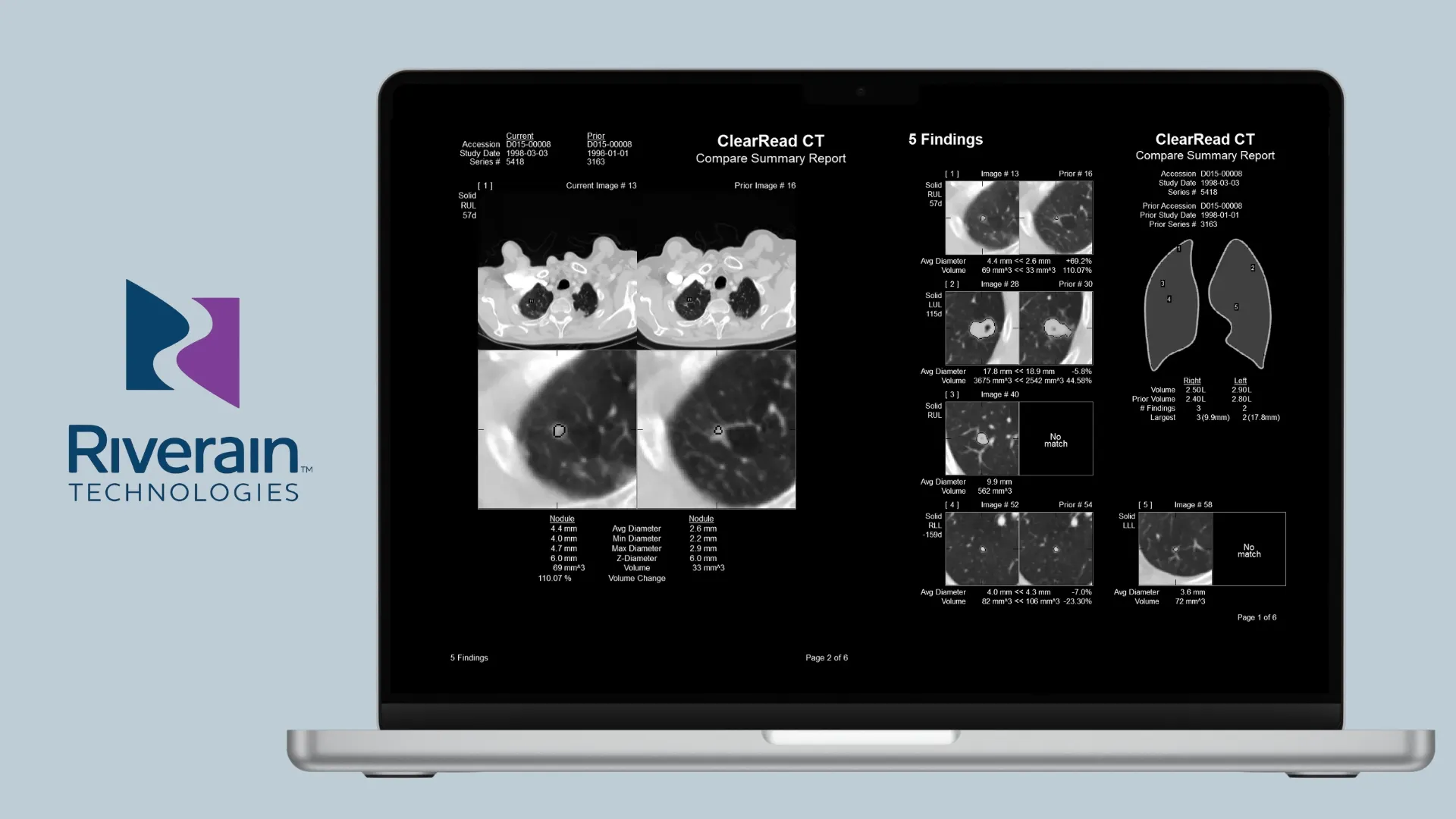
Riverain’s ClearRead CT is an AI-powered solution for thoracic CT interpretation that enhances the detection and quantification of pulmonary nodules by suppressing confounding structures and automating key measurement and comparison tasks. ClearRead CT first generates vessel-suppressed image series to remove vessels and machine noise, providing an unobstructed view of the lung parenchyma that improves conspicuity for solid, part-solid, and ground-glass nodules.
ClearRead CT Detect leverages the vessel-suppressed series to automatically locate, segment, and measure regions of interest, yielding detailed quantitative outputs including nodules’ location, type, volume, maximum, minimum, and average diameters, and depth within the lung field. ClearRead CT Compare extends this capability by automatically matching nodules between current and prior CT studies, enabling efficient visual and volumetric comparisons across timepoints to support longitudinal assessment.
The platform integrates into existing PACS workflows, allowing concurrent viewing of the original and ClearRead-processed series without the need for separate viewers or workstations, and supports structured reporting through integration with reporting tools. ClearRead CT’s architecture includes acquisition normalization technology that enables rapid deployment across CT scanners from different manufacturers and imaging protocols without extensive site-specific tuning.
Infervision – InferRead CT Lung
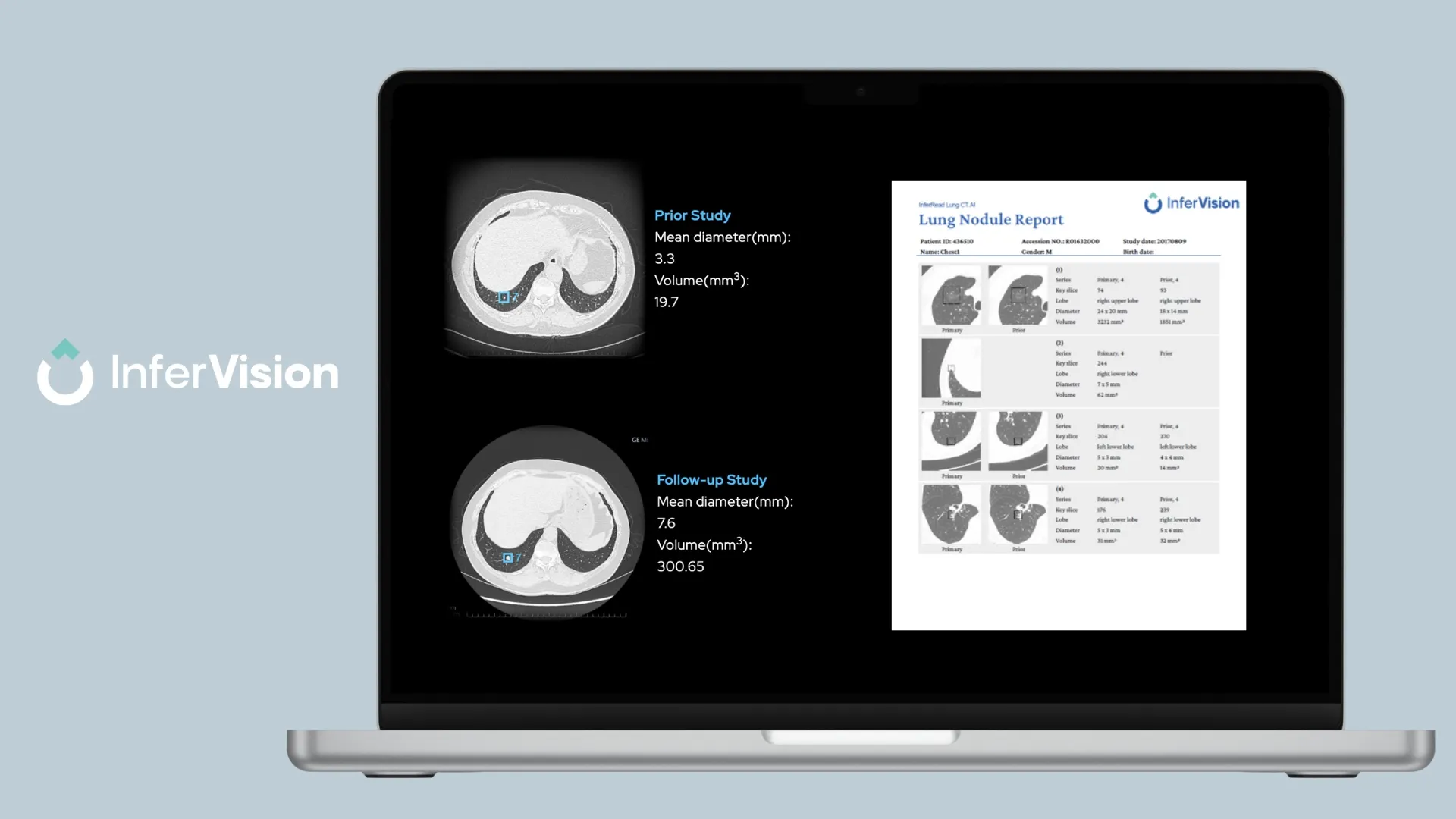
Infervision is a high-tech artificial intelligence company headquartered in Beijing, China, and its product InferRead CT Lung is designed for high sensitivity and comprehensive lung‑health analysis. The vendor reports that AI algorithms detect pure ground‑glass, solid, and mixed nodules and simultaneously measure density and volume in each pulmonary lobe, quantifying lung health parameters for emphysema and interstitial lung disease. It also checks biomarkers for nodule growth, emphysema, and CAC and automatically registers prior and current scans to compute metrics such as volume‑doubling time.
From a workflow perspective, InferRead CT Lung integrates with clinical PACS environments and supports both on-premise and cloud-based deployment options, delivering smart structured reports that populate key findings directly into reporting platforms. The platform’s multi-parameter outputs aim to equip radiologists with consistent, quantifiable data to support interpretation and clinical decision-making across screening and opportunistic cases.
DeepHealth – Chest Suite
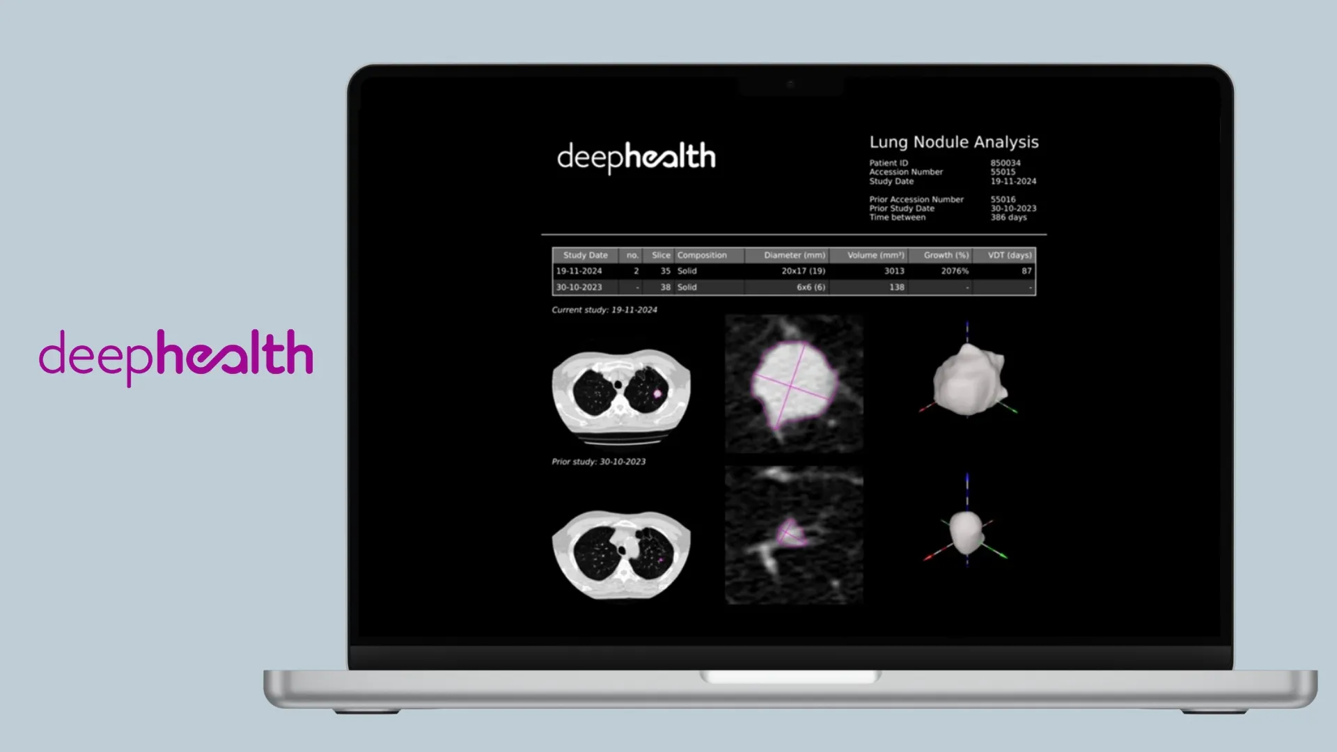
RadNet’s Chest Suite under the DeepHealth brand is a modular set of applications for population‑based lung‑cancer screening and nodule management. At its core is an AI engine that automates the detection and characterisation of pulmonary nodules. The platform provides automated measurements of nodule diameters and volumes over time, which reduces reading time and facilitates longitudinal assessment. It also compares prior and current images to calculate growth percentages and volume‑doubling time. The Chest Suite comprises multiple applications (Veye Lung Nodules, Veye Reporting, DeepHealth Lung AI, DeepHealth Viewer, and HealthCCSng) and positions itself as a cloud‑native platform.
Qure AI – qCT‑Lung & qCT LN Quant
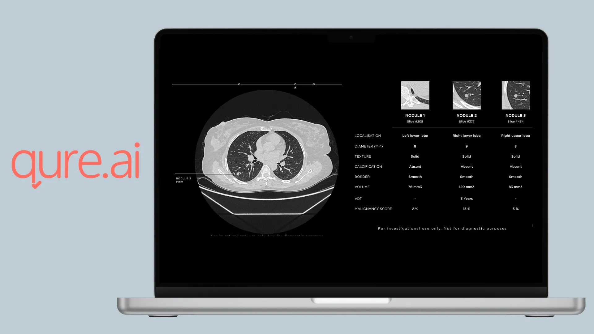
Qure AI is an India-based health-tech company that offers two chest‑CT modules that complement its X‑ray and nodule‑management tools. qCT‑Lung analyzes chest CT scans to detect lung nodules and emphysema and quantifies and tracks nodules over serial exams. The algorithm assesses localisation, spiculation, size, calcification, and texture (solid, sub‑soli,d and ground‑glass), assigns a malignancy risk score, measures volumetry, predicts volume‑doubling time, and can quantify response to treatment. The software pre‑populates reports with suggested follow‑up timelines based on Lung‑RADS and Fleischner guidelines and allows clinicians to adjust which nodules are included.
qCT LN Quant quantifies solid lung nodules on non‑contrast chest CT, as it measures average, short‑axis, long‑axis, and effective diameters, calculates volume‑doubling time, generates Brock malignancy risk scoresand produces 2‑D and 3‑D reconstructions. qCT LN Quant is part of Qure’s lung‑cancer care continuum, which also includes qTrack for nodule management and electronic medical record integration. Together, these tools provide a seamless pipeline from detection to follow‑up and documentation.
Conclusion
For clinical teams evaluating how advanced imaging intelligence can improve prioritisation, longitudinal lesion tracking, and workflow continuity in lung cancer screening, Rayscape offers solutions available today for both Chest X-ray and Lung CT. As our technology evolves toward multi-region analysis and with our new approach to thoraco-abdomino-pelvic oncological analysis, it follows the same principle radiologists use daily: integrating subtle signals across organ systems, reconciling competing findings, and understanding disease as a continuous process rather than isolated observations.
We welcome collaboration with radiologists and institutions committed to advancing oncology imaging practice.
Discover our multiple use cases HERE, or test the solution live through our WEBDEMO.



Discover how Rayscape works
integrate Rayscape AI into
your hospital's infrastructure
and improve both your
daily workflow and patients' experience.
