

Best AI Companies for Chest X‑Ray solutions in 2026
Chest X‑ray remains one of the world’s most common imaging tests, yet reviewing hundreds of radiographs per day makes it easy to miss subtle lung findings. Forty percent of all radiology images in hospitals are chest radiographs, and interpreting them correctly is challenging because pathologies often overlap and the anatomy of the chest is complex.
Unlike cross‑sectional CT scanning, chest radiography compresses three‑dimensional anatomy into a two‑dimensional image, complicating detection of small nodules, bone lesions, or early lung disease. Radiologists, therefore, rely on computer‑aided detection (CAD) to highlight suspicious regions, quantify thoracic metrics, compare exams over time, and prepare structured reports that reduce clerical effort.
In recent years, AI‑driven imaging vendors have matured from simple anomaly spotters into comprehensive workflow tools that classify dozens of pathologies, integrate seamlessly with PACS/RIS, and perform semi‑automated reporting. This article reviews seven commercially available solutions for chest radiography in 2026, focusing on the features that distinguish each company.
Rayscape - the Radiology AI for Chest X-ray
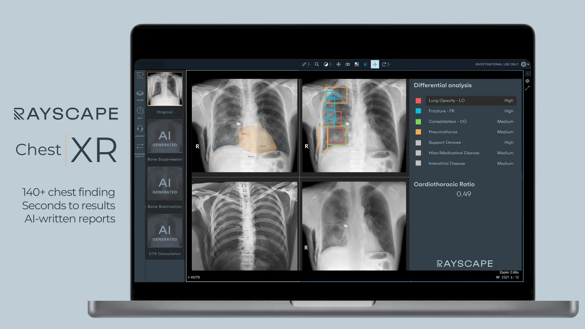
Rayscape CXR is engineered as a clinical infrastructure, not a narrow algorithm. The solution operates as a diagnostic support layer across the entire chest radiography workflow, transforming raw images into structured, longitudinally coherent clinical information that scales across low or high-volume environments. The system is designed to support radiologists with organized, reproducible outputs rather than isolated algorithmic flags, reinforcing consistency from image acquisition through report generation. The AI developed by the Rayscape team helps with:
- Pathology identification and localization: The platform identifies and classifies more than 140 radiographic findings across 17 major pathology superclasses, including consolidation, pneumothorax, pleural effusion, nodules and masses, fractures, atelectasis, interstitial patterns, and device-related findings. Each suspected abnormality is localized with bounding boxes and compiled into a structured findings list, allowing rapid anatomical orientation and prioritization.
- AI-generated advanced visualizations: Bone-suppression and subtraction reconstructions are produced alongside the original radiograph, improving visibility of soft-tissue structures and subtle osseous abnormalities that may be obscured by overlapping anatomy.
- Cardiothoracic ratio analysis: The system automatically calculates the cardiothoracic ratio and provides a visual representation directly on the study, embedding quantitative cardiac assessment into routine chest interpretation.
- Customisable clinical interface: The user environment is fully configurable, allowing institutions to align outputs, thresholds, and visualization preferences with local workflow, reporting standards, and clinical priorities.
- LLM-ready structured reporting: Detected findings can be consolidated by an integrated reporting engine that converts AI outputs into a coherent draft radiology report. This produces structured, narrative-ready documentation for radiologist review and validation, reducing manual transcription while preserving physician oversight.
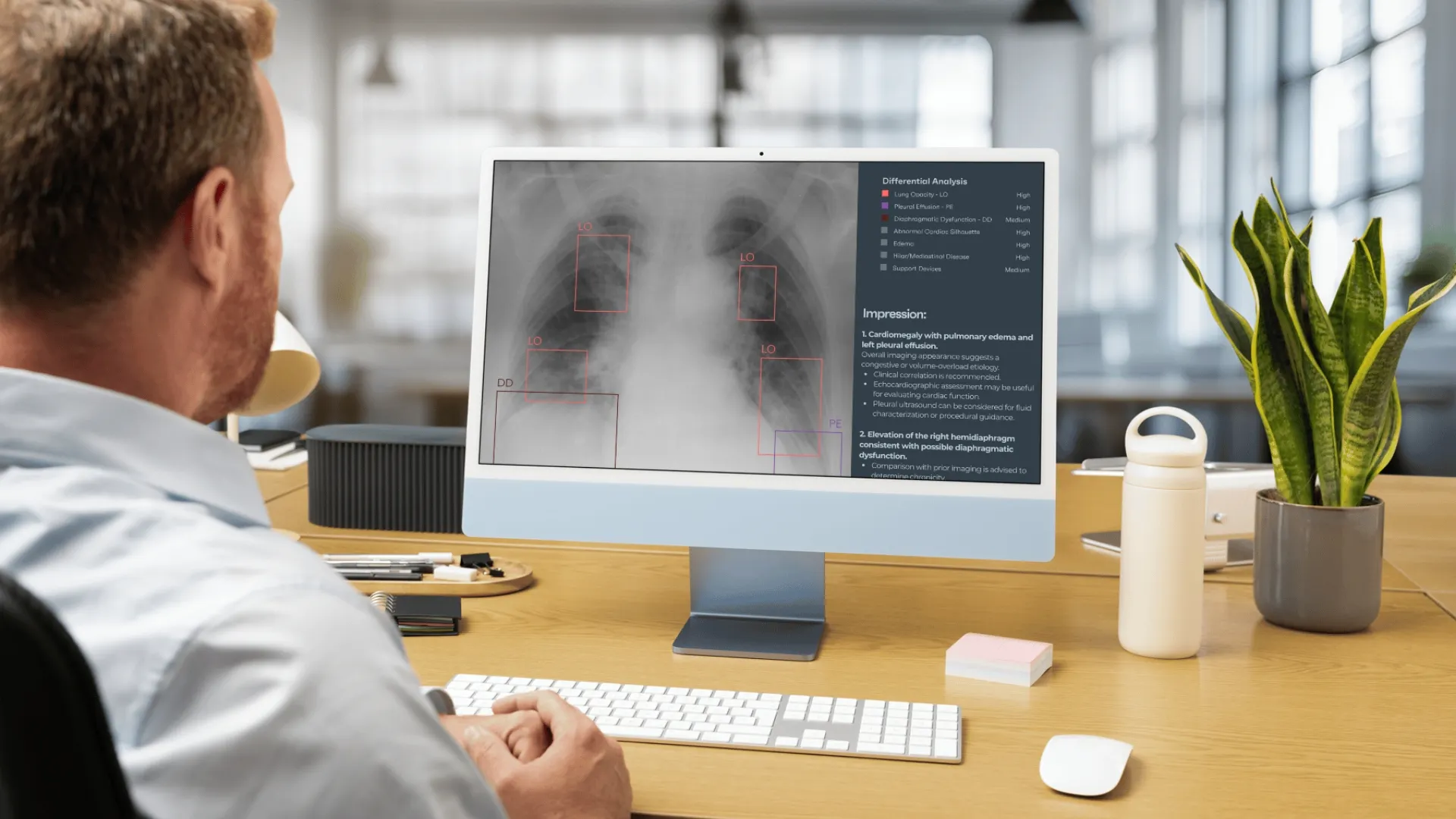
Deployment follows a plug-and-scan model designed for operational environments. The solution is CE-marked, externally evaluated, and the system integrates directly into on-premise or cloud PACS infrastructure, returning annotated images and structured outputs within the native radiology viewer. No parallel workspace. No workflow disruption. Intelligence remains embedded where interpretation already occurs. The architecture extends beyond CXR, linking with complementary CT modules that support lung cancer screening pathways. This cross-modality enables unified thoracic imaging programs spanning emergency departments, ICUs, outpatient centers, teleradiology companies, TB screening initiatives, and population-scale programs where consistency, volume handling, and longitudinal tracking define performance.
Gleamer - ChestView
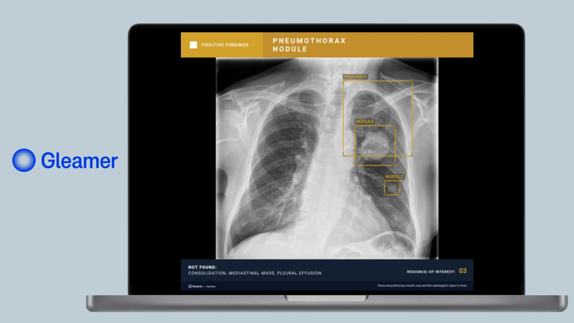
Gleamer’s ChestView acts as a second reader for 5 high‑risk abnormalities: pneumothorax, consolidation, pulmonary nodule, mediastinal or hilar mass, and pleural effusion. The algorithm automatically measures the cardiothoracic ratio to assist in detecting cardiomegaly. ChestView was co‑developed with Assistance Publique–Hôpitaux de Paris and is underpinned by a robust cross‑annotated database that has been validated and deployed at scale. The AI findings can be summarized into structured elements that integrate into PACS/RIS reporting environments, turning detections into usable documentation. ChestView targeted detection of the most urgent thoracic pathologies means it complements, rather than replaces, full‑coverage solutions.
AZmed - AZchest
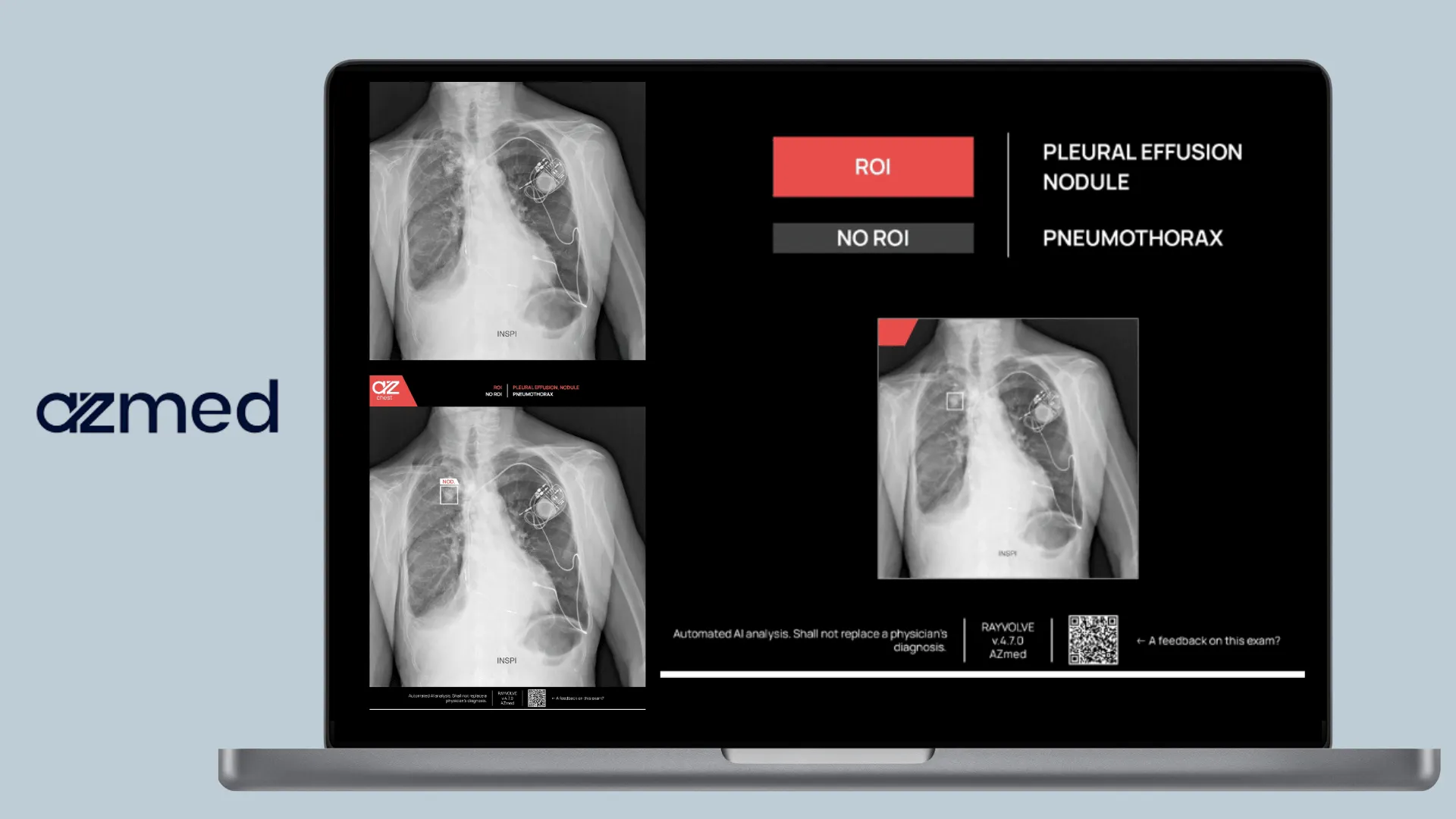
AZmed’s AZchest (formerly Rayvolve) uses deep learning to screen frontal and lateral chest radiographs for 7 key lung and heart conditions: lung nodules, rib fractures, cardiomegaly, consolidation, pleural effusion, pneumothorax, and pulmonary edema. AZchest is built as a background clinical support tool for chest radiography, functioning as an automated second reader within routine workflows. Once connected to PACS, the system analyzes standard chest X-rays to flag the defined set of high-impact findings, supporting prioritization without altering the radiologist’s primary reading process. Detected regions are highlighted with bounding boxes, and results are summarized into structured outputs that support triage and rapid case orientation.
Oxipit - CXR Suite
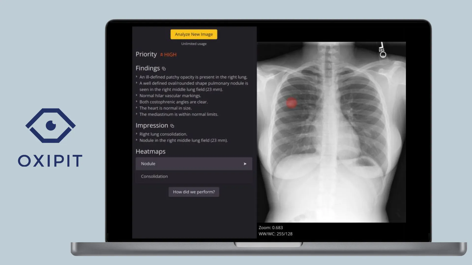
Oxipit is a Lithuanian-based radiology AI developer. Their product, CXR Suite, encompasses three modules—ChestLink, ChestEye, and Quality—that together aim to automate normal‑case reporting. The platform detects 75 findings, ranging from lines and tubes to lung opacities, fractures, masses, pneumothorax, and tuberculosis. It can identify “normal” studies and automatically issue standard reports, freeing radiologists from routine follow‑ups. A real‑time quality module continuously checks AI‑negative studies for missed abnormalities. Oxipit emphasises scalability as the system can handle workloads from 5,000 to more than 500,000 radiographs per year and has minimal IT requirements for integration. A web‑based analytics dashboard offers triage oversight, performance metrics, and continuous updates, while the suite’s auto-prioritisation ensures that suspicious cases rise to the top of the worklist.
Harrison.ai - CXR
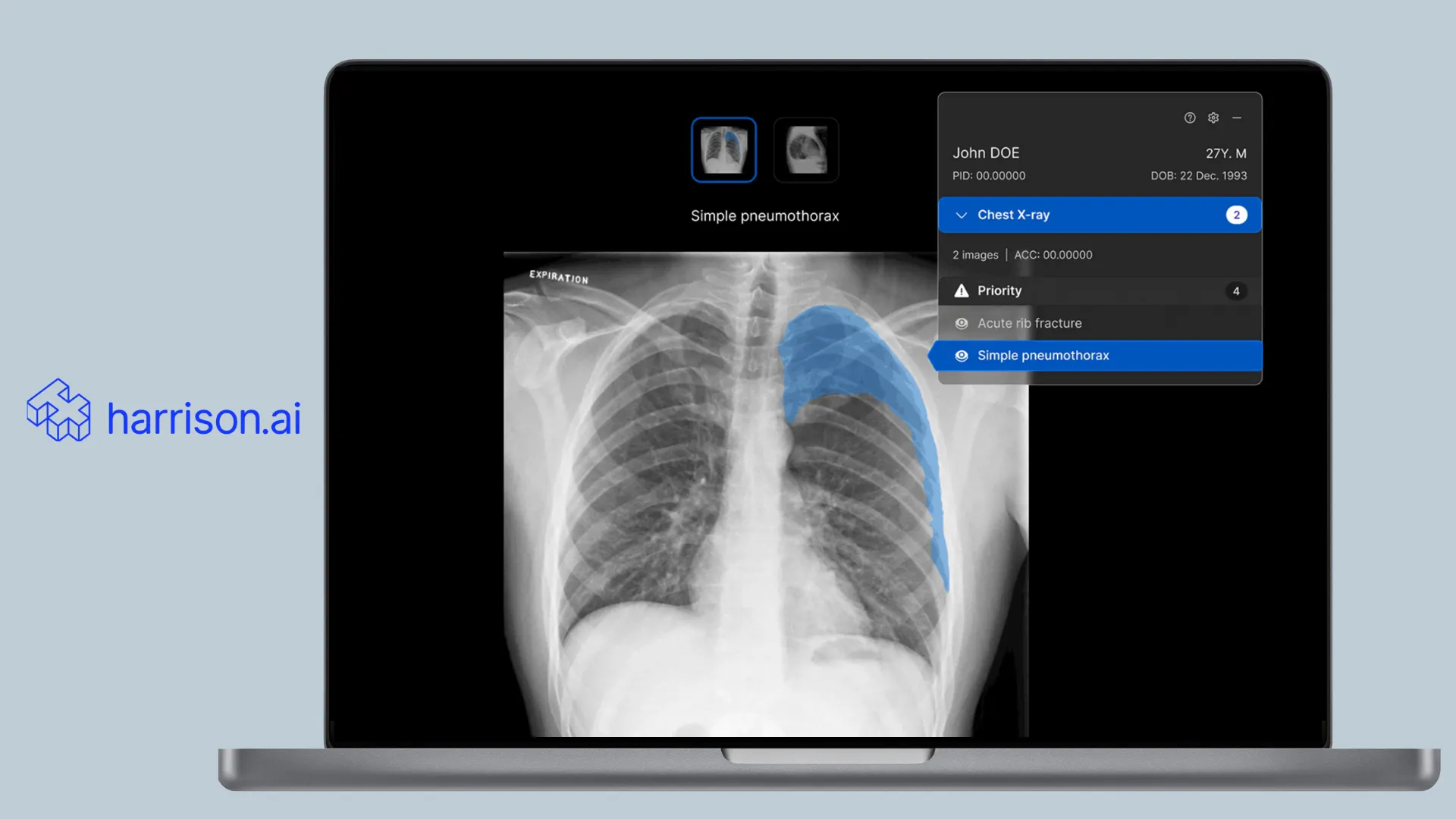
Harrison.ai’s chest‑radiography solution is positioned as a workflow‑integrated platform that detects up to 124 findings. The user interface overlays colour‑coded heat‑maps onto the radiograph and displays a resizable list of detected findings, each accompanied by a confidence bar and bounding box. Radiologists can customise detection thresholds, hide or reveal specific findings, and toggle multi‑image views such as PA and lateral projections. From a deployment standpoint, the solution is CE marked and offers a no‑click viewer that inserts AI results directly into existing PACS worklists. The combination of granular classification, explainable outputs, and seamless integration makes Harrison.ai a good candidate for health systems seeking AI assistance without sacrificing user control.
Lunit - INSIGHT CXR
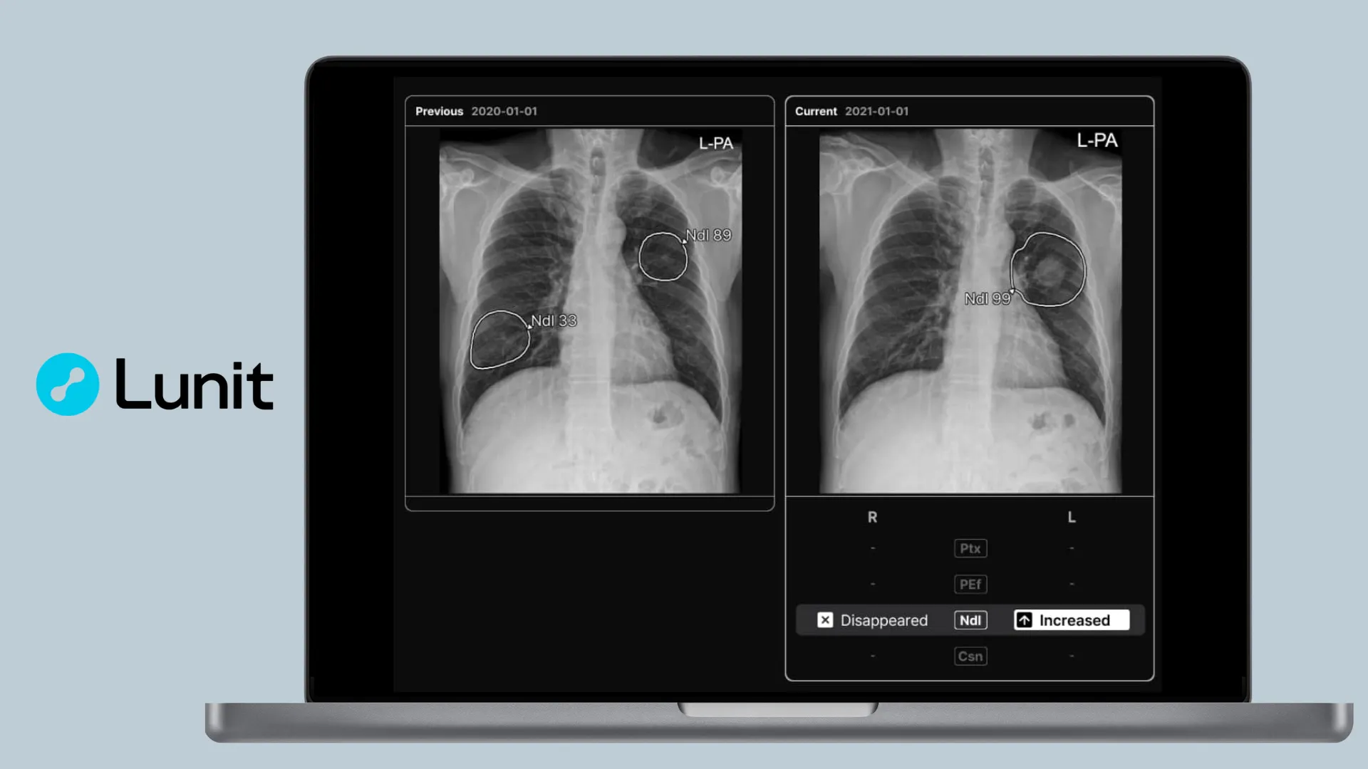
Lunit, a South Korean AI oncology imaging software, is the provider of INSIGHT CXR. The software employs a DualScan architecture comprising two AI engines: one detects abnormalities and the other flags normal exams, thereby reducing backlogs and expediting normal‑case reporting. The engine highlights 11 major thoracic abnormalities—including lung nodules, consolidation, pneumothorax, pneumoperitoneum, pleural effusion, mediastinal widening, cardiomegaly, calcification, fibrosis, atelectasis, and acute bone fracture—and supports tuberculosis screening. It generates normal reports when no pathology is found.
Riverain - ClearRead X-ray
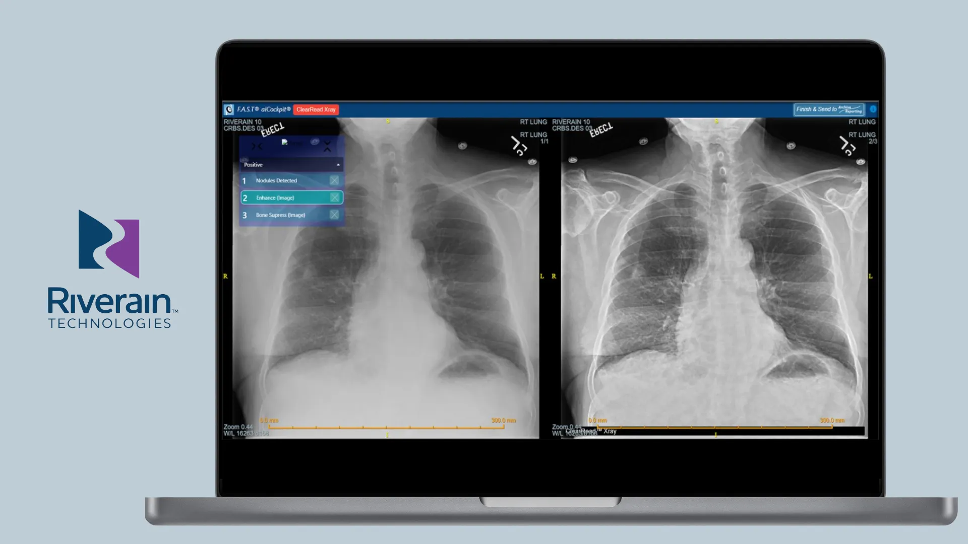
Riverain’s ClearRead X-ray is a suite of applications that enhance chest‑radiograph interpretation. Bone Suppress creates a bone‑suppressed series to improve soft‑tissue visibility, which helps radiologists discover nodules previously obscured by the ribs or clavicles. Detect automatically identifies and marks pulmonary nodules (particularly those 9–30 mm in size) on the bone‑suppressed image, reducing false positives. Enhance highlights lines and tubes to improve visibility in portable radiographs, and can be toggled on or off to speed up interpretation. Riverain also offers Pneumothorax and Compare applications: the pneumothorax module triages potential collapses, while Compare automatically matches nodules across current and prior radiographs for longitudinal assessment. All modules integrate into existing PACS viewers and work across imaging systems without additional hardware or radiation exposure.
Conclusion
While numbers such as sensitivity or specificity often dominate discussions about AI performance, they seldom capture how these solutions actually change radiologists’ daily practice. For teams that want to advance their imaging workflows, Rayscape provides operational imaging intelligence for Chest X-ray and Lung CT, supporting prioritization, longitudinal lesion tracking, longitudinal assesment and multimodality across care pathways. As our technology evolves toward multi-region analysis and with our new approach to thoraco-abdomino-pelvic AI detections, it follows the same principle radiologists use daily: integrating subtle signals across organ systems, reconciling competing findings, and understanding disease as a continuous process rather than isolated observations.
We welcome collaboration with radiologists and institutions committed to advancing their imaging practice.
Discover our multiple use cases HERE, or test the solution live through our WEBDEMO.



Discover how Rayscape works
integrate Rayscape AI into
your hospital's infrastructure
and improve both your
daily workflow and patients' experience.
