Introduction
A 2026 open-access study published in Current Health Sciences Journal evaluated the impact of AI assistance on radiologist performance for pulmonary nodule detection on chest CT. The study was conducted by researchers from the University of Medicine and Pharmacy of Craiova and Craiova County Emergency Clinical Hospital and specifically evaluated Rayscape as the AI-based lung nodule assessment system.
Pulmonary nodules represent one of the most important imaging findings in the early detection of lung cancer. Although CT provides substantially greater sensitivity than conventional chest radiography, interpretation remains dependent on the radiologist reviewing a large number of images, often in examinations containing multiple or subtle nodules.
Observer-related factors including fatigue, distraction, lesion appearance, anatomical location and the number of findings present in an examination can contribute to missed nodules and variability between readers. The study therefore investigated whether adding an AI system as a second reader could reduce missed findings and improve agreement with an expert reference standard.
The primary objective was to compare three approaches:
a) radiologist-only interpretation;
b) radiologist interpretation assisted by Rayscape;
c) the AI system operating independently.
Performance was evaluated against an expert reference standard, with particular attention to detected and missed nodules, false-positive findings, diagnostic error, sensitivity, specificity and agreement with the reference reader.
Materials and Methods
The retrospective study included 300 patients aged 40–80 years who underwent chest CT examinations during September and October 2024 at the Medical Imaging Department of the University of Medicine and Pharmacy of Craiova. The cohort consisted of 137 women and 163 men and included both patients with known malignancies and individuals without significant previous medical history.
Patients with previous lung surgery, incomplete diagnostic reports or significant respiratory motion or beam-hardening artefacts that could interfere with interpretation were excluded. All examinations were acquired using a single Siemens multidetector CT system. Contrast-enhanced chest CT was performed during a single inspiratory breath-hold using 120 kV acquisition and 1.5 mm slice thickness.
The reading workflow involved several stages. An initial report was generated by a junior radiologist and reviewed by a board-certified radiologist without AI assistance. A different radiologist, blinded to the original report, subsequently reviewed the examinations using Rayscape. Finally, the findings from the radiologist-only reading, AI-assisted reading and standalone AI report were recorded and compared with a reference assessment established by a senior board-certified radiologist experienced in thoracic imaging.
The evaluated AI system was the commercially available lung nodule assessment solution developed under the Rayscape trade name. CT examinations were transferred directly from the scanner to the AI server for automated processing.
The software automatically detected pulmonary nodules and presented them on the radiologist's workstation using bounding boxes together with quantitative and structured information including:
a) anatomical location;
b) nodule diameter;
c) nodule volume;
d) imaging characteristics;
e) prioritisation of suspicious findings;
f) follow-up recommendations based on Fleischner Society criteria.
Reported processing time was approximately 10–20 minutes per examination. Statistical analysis included paired-samples t-tests, Wilcoxon signed-rank testing, Pearson correlation, linear regression and mean absolute error (MAE). Sensitivity and specificity were also calculated, with statistical significance defined as p < 0.05.
Results
1. Reduction in missed pulmonary nodules
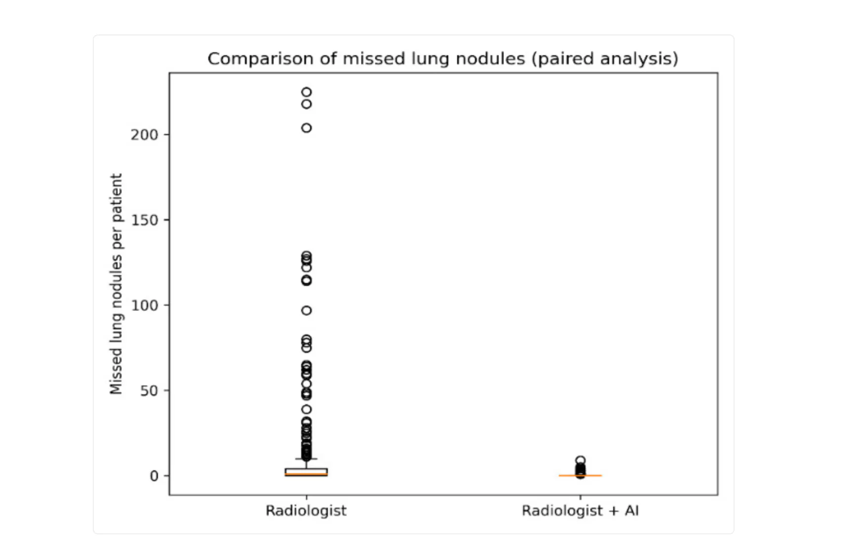
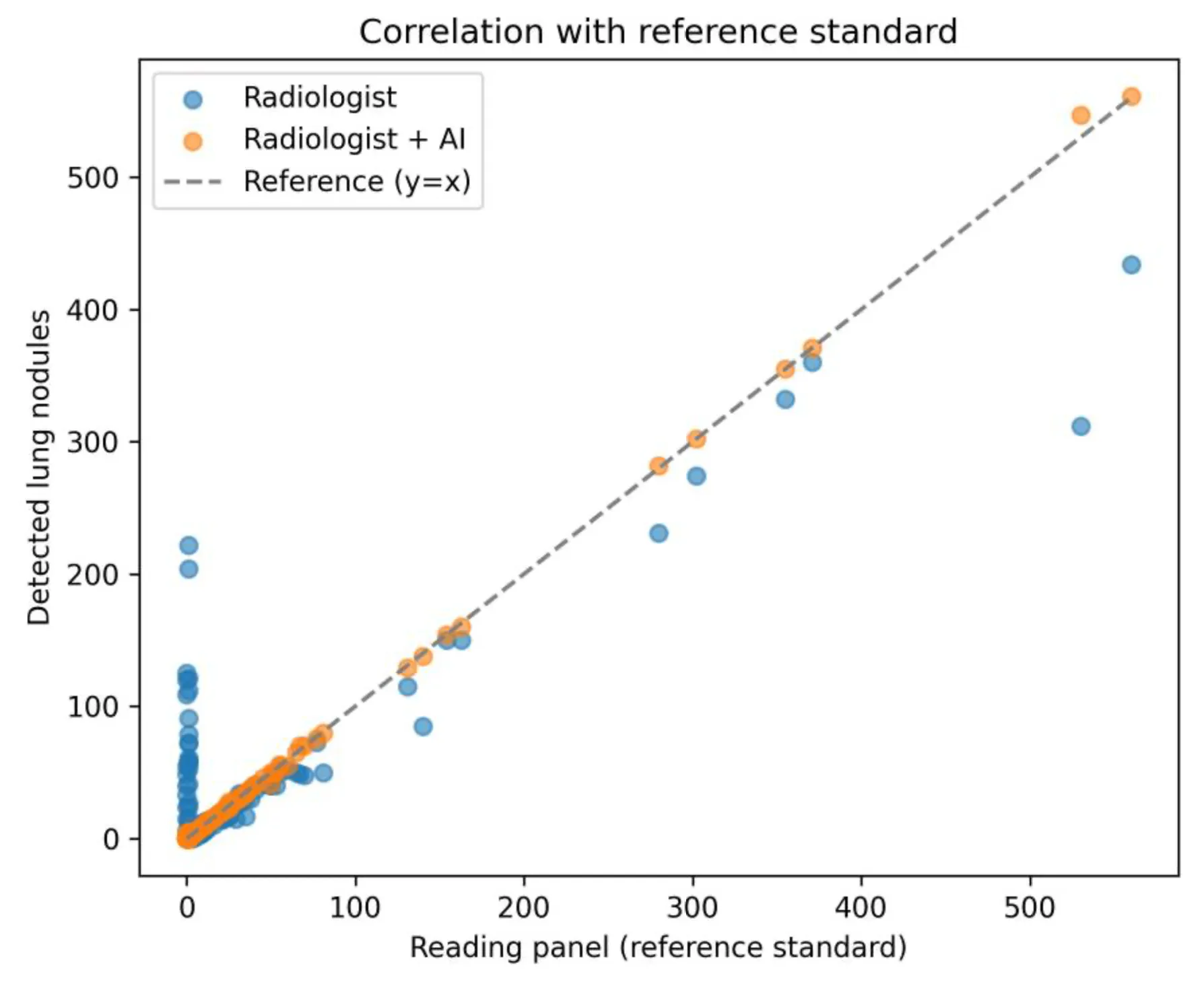
AI assistance produced a statistically significant reduction in the number of nodules missed by radiologists. The difference between radiologist-only and AI-assisted readings was significant with t = 5.71 and p < 0.001, and the distribution of missed nodules was also less variable when Rayscape was used.
The study reported the following overall number of detected nodules:
a) Radiologist alone: 6,760
b) Radiologist + Rayscape: 5,811
c) Rayscape standalone: 7,959
d) Expert reference panel: 5,783
The AI-assisted radiologist therefore produced a total count substantially closer to the expert reference value than either the unassisted radiologist or the standalone AI output.
2. Agreement with the expert reference standard
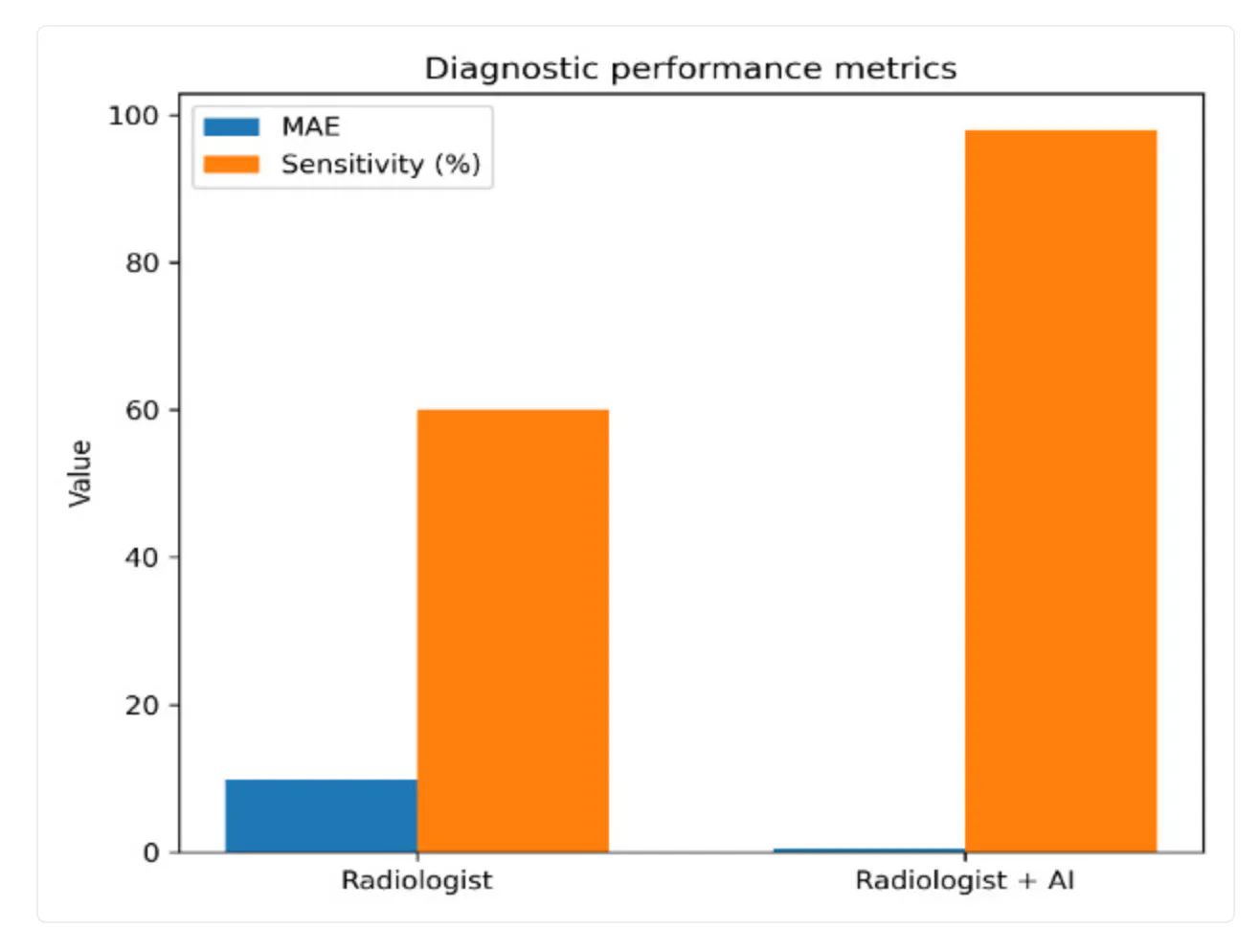
The radiologist-only interpretation demonstrated a strong correlation with the expert reference standard at r = 0.861. With Rayscape assistance, agreement increased to an almost perfect correlation of approximately r = 1.00. Diagnostic error showed a similarly substantial improvement. The study reported a reduction in mean absolute error from 9.78 to 0.46 when the radiologist used AI assistance. The difference in diagnostic errors was statistically significant on Wilcoxon signed-rank testing (p < 0.001), indicating that AI-assisted results were consistently closer to the expert reference assessment.
3. False-negative and false-positive findings
One of the clearest findings of the study was the reduction in both missed nodules and false-positive findings when AI was combined with radiologist interpretation. The number of reported false negatives therefore decreased from 3,083 to 145 with AI-assisted interpretation, while false positives decreased from 2,073 to 59.
The standalone AI system also demonstrated strong detection performance, but produced more false-positive and false-negative findings than the combined radiologist + AI workflow.
This distinction is important: the strongest performance in the study was achieved not by replacing the radiologist, but by combining the radiologist's interpretation with AI assistance.
4. Sensitivity and specificity
AI assistance substantially improved the reported diagnostic performance:
a) Sensitivity: increased from 60% to 98%
b) Specificity: increased from 69% to 99%
c) Standalone AI sensitivity: 95%
d) Standalone AI specificity: 94%
The combination of radiologist interpretation and Rayscape therefore achieved the highest reported sensitivity and specificity among the evaluated approaches.
5. Workflow implications
The study also highlights the workload associated with examinations containing large numbers of pulmonary nodules. According to the authors, visual evaluation could require only several minutes in examinations containing fewer than 10 nodules but could reach 45–50 minutes in patients with more than 100 nodules.
AI-assisted reading facilitated faster identification and evaluation of these findings, particularly in high-burden examinations. However, the study does not report a controlled radiologist reading-time comparison between assisted and unassisted workflows, so the workflow benefit should be interpreted as an observed advantage rather than a formally quantified time-saving endpoint.
Discussion
The study demonstrates a clear improvement in pulmonary nodule detection when Rayscape is used as a second reader alongside the radiologist. The most significant result is the reduction in missed nodules. False negatives decreased from 3,083 in the radiologist-only assessment to 145 with AI assistance, accompanied by an increase in sensitivity from 60% to 98%.
Conclusion
The study provides clinical evidence that Rayscape-assisted chest CT interpretation can substantially improve pulmonary nodule detection compared with radiologist-only reading.
Across 300 clinical examinations, AI-assisted interpretation was associated with fewer missed nodules, substantially lower diagnostic error, increased sensitivity and specificity, and almost perfect agreement with an expert reference standard. Importantly, the results favour a radiologist + AI model rather than standalone AI. The strongest diagnostic performance was achieved when Rayscape functioned as a second reader, combining automated detection and quantitative analysis with the radiologist's clinical judgement.
Within the setting evaluated by the study, Rayscape increased sensitivity from 60% to 98%, reduced false negatives from 3,083 to 145, and reduced mean absolute error from 9.78 to 0.46. These results support the clinical value of AI-assisted reading for more systematic and consistent pulmonary nodule assessment in routine chest CT.
LUNG CANCER SCREENING WITH RAYSCAPE
Rayscape Lung CT supports radiologists in detecting and assessing pulmonary nodules by automatically highlighting findings and providing structured measurements such as diameter and volume directly within the CT workflow.
Designed to act as a second reader, it helps surface relevant findings, support more consistent assessment, and reduce manual work. The solution integrates into existing hospital and PACS infrastructure and can support lung cancer screening, incidental nodule detection, routine chest CT, and longitudinal follow-up.
For scientific inquiries, validation details, and integration into lung cancer screening workflows, contact us and learn more!