

Pulmonary edema detection with Rayscape AI
Pulmonary edema, also known as pulmonary congestion, is excessive liquid accumulation in the tissue and air spaces (usually alveoli) of the lungs. It causes poor oxygen delivery, which can lead to hypoxemia and respiratory failure. It occurs when the left ventricle of the heart fails to appropriately remove oxygenated blood from the pulmonary circulation (cardiogenic pulmonary edema), or when lung tissue or blood vessels are injured directly (non-cardiogenic pulmonary edema).
While typically being caused by a dysfunction of the left ventricular muscle, fluid can build up on its own due to lung injury. External traumas or lesions caused by elevated pressures inside the respiratory system can cause this type of edema. Pulmonary liquid can occur when average pulmonary pressure increases from the usual norm of 15 mmHg to above 25 mmHg. Pulmonary edema can be classified into two types: cardiogenic and non-cardiogenic.
HOW DOES PULMONARY EDEMA OCCUR?

Congestive heart failure is the most common cause for cardiogenic type. When the heart can no longer pump blood properly throughout the body, it is called heart failure. In patients with left sided heart failure, the left ventricle becomes unhealthy and can’t contract effectively. Because of this, blood starts to accumulate in the left atrium and from here it follows a backwards path towards pulmonary veins. This produces a build-up of pressure in the lungs vascularization (pulmonary hypertension), causing them to leak fluid into the interstitial space.
Another cardiogenic cause is the severe systemic hypertension, specifically blood pressure that is greater than 180/110 mmHg systolic/diastolic. In this situation the left ventricle is healthy but can’t effectively pump blood in the circulatory system due to the resistance in front of it.

Non-cardiogenic causes of pulmonary edema include pulmonary infections, inhalation of toxic substances or trauma to the chest. Pulmonary embolism, high altitude or seizures can also be the causing agent. All of this can create direct injury to the alveoli, (tiny balloon-shaped structures at the end of the respiratory tract ramification). When this happens, there is usually an inflammatory process that makes nearby capillaries more permeable. As a result, proteins and fluid enter the interstitial space.
Another category of non-cardiogenic cause is the low oncotic affections (stage where the pressure that keeps the fluid in the vascular space is too low). The most common pathologies with low oncotic pressure are malnutrition, liver failure or nephrotic syndrome.
WHAT ARE THE SYMPTOMS?
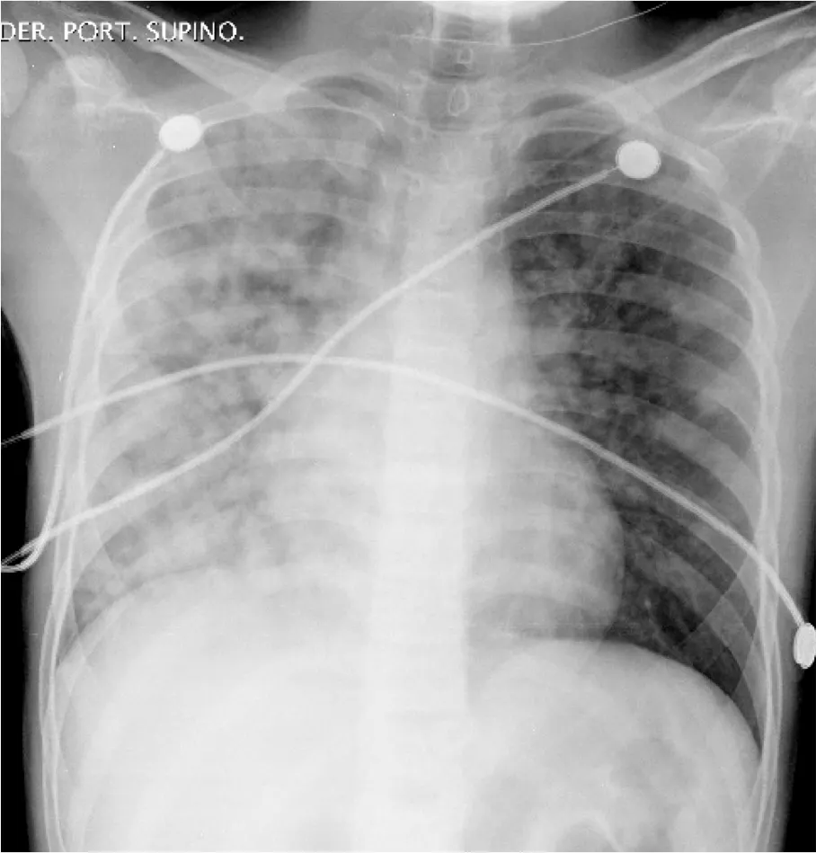
Acute pulmonary edema can appear suddenly and can be life-threatening. The following are the most prevalent symptoms:
- Sudden shortness of breath, especially after intense activity or while lying down
- Anxiety
- Palpitations and sweating
- Wheezing and gasping for air
- Coughing up pink, frothy spit
- Cyanosis
- Dizziness and weakness
When the pulmonary fluid builds up over time, it’s called chronic pulmonary edema. In this case the patient may have the following symptoms:
- Tiredness
- Weight gain
- Swollen legs and feet
- Trouble breathing when lying down
- Waking up at night frequently
The chest radiograph is still the most practicable and appropriate way to assess and quantify pulmonary edema. On a chest X-ray, the following features are useful for quantifying pulmonary edema:
- Pulmonary venous enlargement in the upper lobes
- Higher cardiothoracic ratio
- Perihilar haze and peribronchial cuffing
- Septal lines (Kerley)
- Pleural effusions and fluid in interlobar fissures
- Thickening of interlobar fissures
- Air space opacification
Chest computed tomography (CT) has also been used in the noninvasive examination of pulmonary edema, with the added benefit of being able to quantify lung density as a reference for changes in lung water content. . On a chest CT, the following features are useful for quantifying pulmonary edema:
- ground glass opacification (increased lung opacity through which vessels and bronchial structures may still be seen.
- bronchovascular bundle thickening (due to increased vascular diameter and/or peribronchovascular thickening)
- interlobular septal thickening
The pulmonary edema should be detected as soon as feasible because of the variety of forms it can take and the therapeutic urgency it poses. It is characterized by dyspnea and hypoxia secondary to fluid accumulation in the lungs which impairs gas exchange and lung compliance. Because of this, the oxygenation and ventilation are insufficient to meet the metabolic demands of the body.
AI FOR FASTER DIAGNOSIS
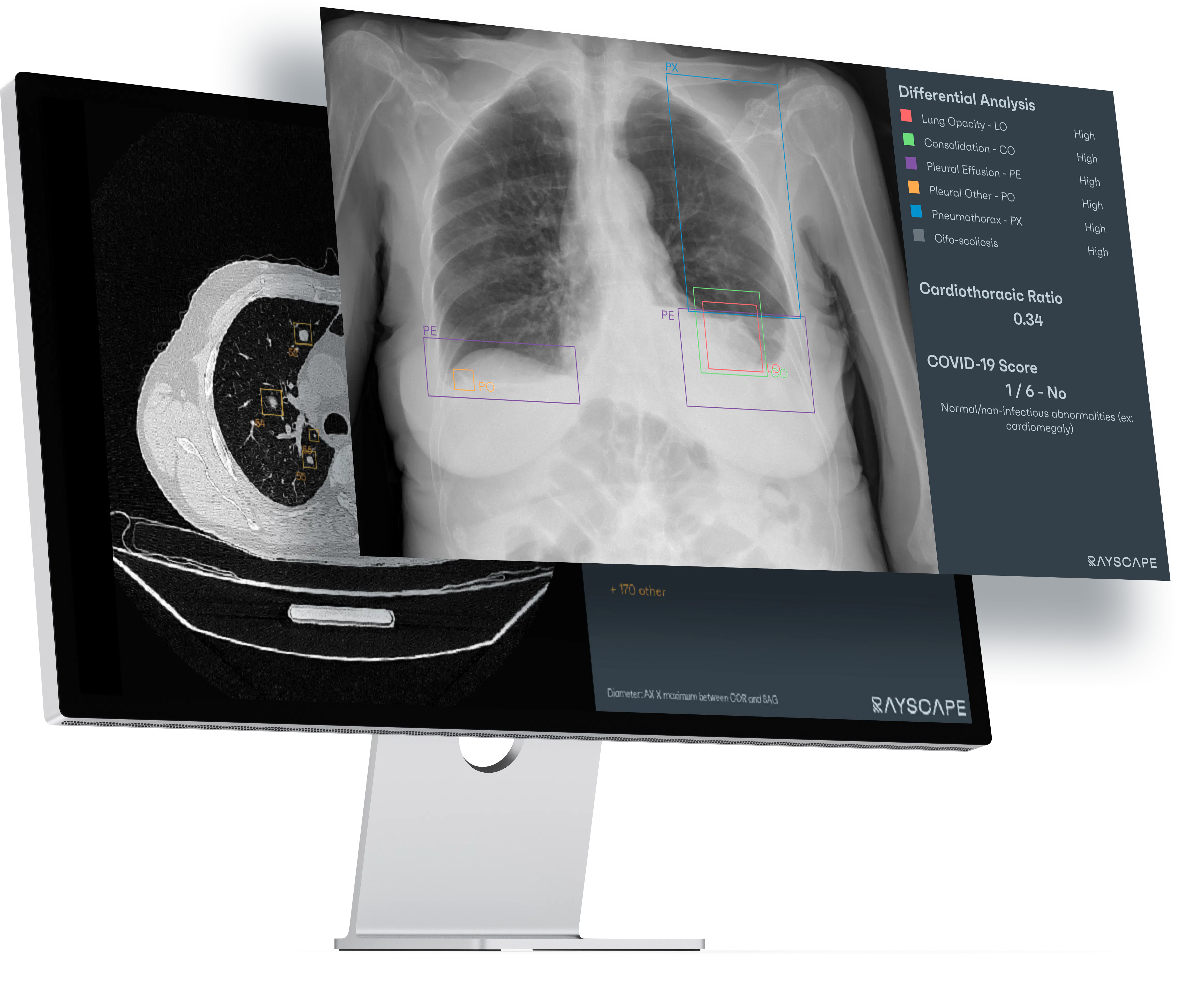
Our software aids professionally qualified radiologists and clinicians when diagnosing the affection on a chest radiography, by detecting abnormal pathologies.
We help obtaining a faster diagnosis by providing deep neural networks trained on a large collection of chest radiographies. Rayscape contains 3 different algorithms that serve different functions:
1) A chest X-ray screening algorithm that is specifically trained and optimized to differentiate between normal and abnormal chest radiographies, regardless of the pathology encountered in the medical image. Its main use is to help radiologists in prioritizing their workload.
2) A chest X-ray pathology detection algorithm that can detect over 100 pathologies that are classified in 17 different classes of pathologies that include most of the existing chest radiological findings. It also generates a bounding box for each class of pathologies detected on the x-rays.
This way it emphasizes the areas that are considered anomalous by the algorithm, as well as an automated calculation of the cardio-thorax index. This helps identifying missed pathologies by prompting the user to not skip underreported medical findings.
3) A bone subtraction and suppression algorithm that generates two additional x-ray visualizations of the original chest radiography. The bone suppressed visualization emphasizes the bone tissue in the image, making it much easier for our users to spot fractures or calcifications. The bone subtracted one only shows soft tissue, increasing the visibility of lesions that hide behind bones.
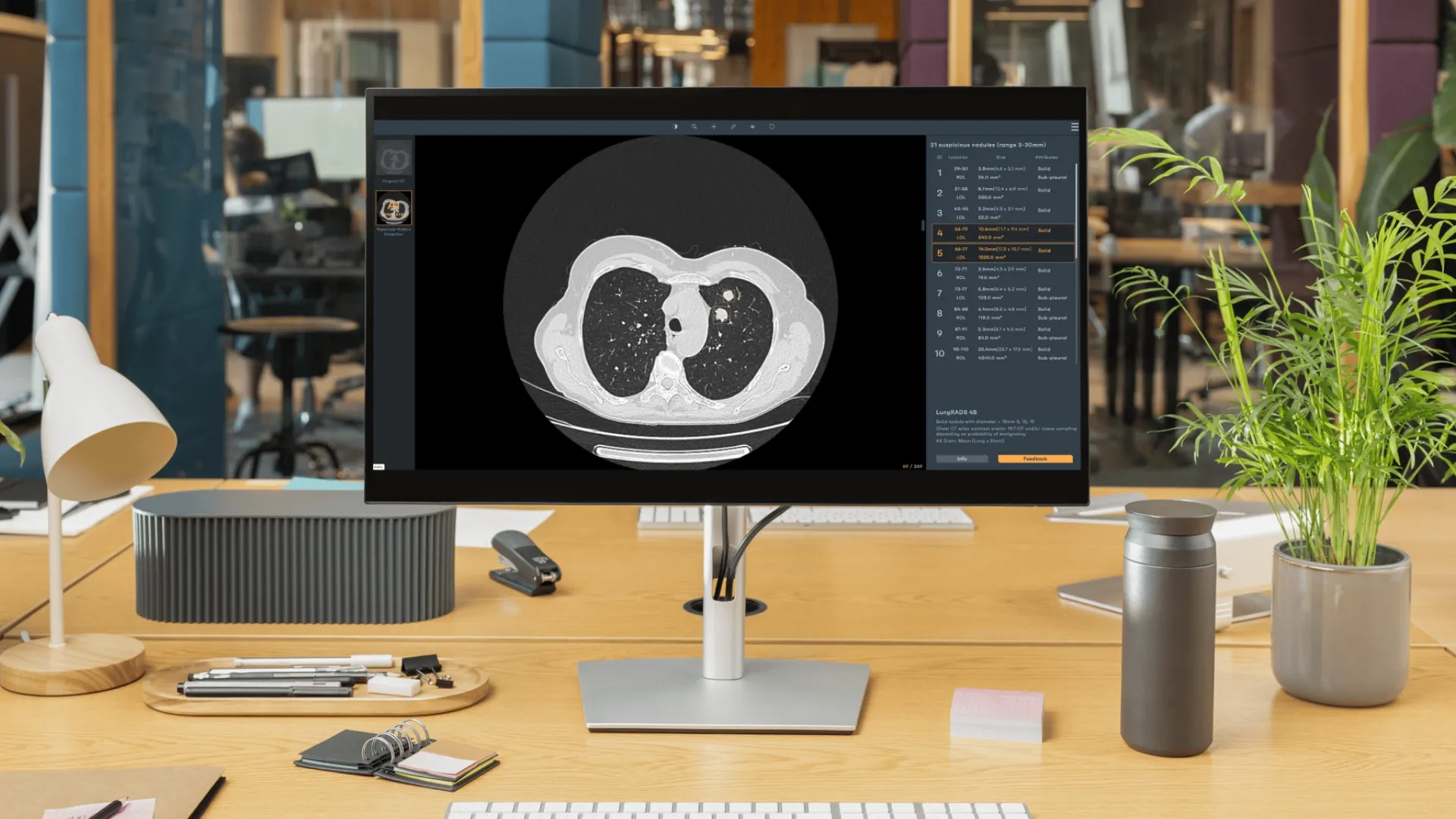
Our seamless integration allows the user to view the analyzed images directly in their PACS screen, without the use of an external application. This way, we provide radiologists with a method to prioritize and triage patients better, while making them more effective.
Using Rayscape, a doctor can decrease the total time to diagnose each patient, as well as increase the precision of X-ray interpretations, especially during night shifts or during periods of time with increased workloads. Because of this, cases of pulmonary edema can be identified more quickly and be treated in a timely manner.
Our team is dedicated to creating the best digital radiology system for medical imaging interpretation. All of them powered by AI.


Discover how Rayscape works
integrate Rayscape AI into
your hospital's infrastructure
and improve both your
daily workflow and patients' experience.
