

AI Oncology Imaging: from radiology detection to context & prioritization
Radiology has become the central nervous system of modern oncology. Each cancer journey is documented through waves of imaging studies: initial AI radiology oncology imaging for detection, diagnosis, serial CT scan analysis to evaluate response, and periodic surveillance to hunt for recurrence. Current advances in AI thorax‑abdomen‑pelvis computed tomography have given machines the ability to spot tumours, nodules and metastases, yet radiologists and oncologists are still drowning in data.
A typical cancer workup produces hundreds or even thousands of cross‑sectional images spanning the lungs, liver, lymph nodes, bones and pelvic organs. As technology advances, detection becomes less of a triumph and more of a footnote. The bottleneck moves to the part no algorithm can solve by brute force alone: deciding what matters first, understanding what a finding means in the context of the patient’s anatomy and timeline, and surfacing the right signal before the plot moves on. This article explores why detection is now table stakes and what must come next for oncology cancer screening AI imaging apps.
The shifting bottleneck in oncology imaging
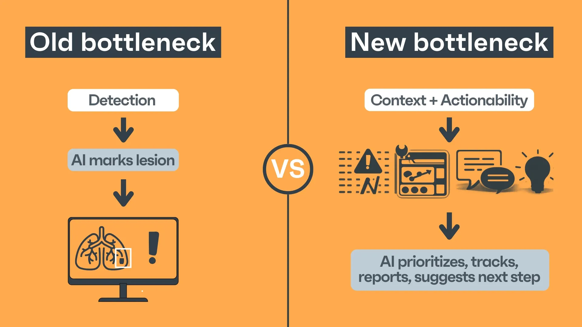
For years, the primary challenge in computational radiology was enabling machines to identify abnormalities with high sensitivity and specificity. That challenge is still present, but progress is being made by AI vendors in the field. Deep convolutional networks, transformers and ensemble models routinely approach expert‑level performance at tasks such as lung nodule detection, liver segmentation or pelvic bone fracture identification. Vendors and open‑source projects offer off‑the‑shelf detectors for the most common pathologies, and yet, a comprehensive solution for oncology is still not available. In routine practice, the false‑positive and false‑negative rates of these systems are low enough that their incremental gains over radiologists’ detection accuracy are marginal.
Meanwhile, imaging volumes have exploded. Screening programmes, personalised medicine and improved survival have led to an avalanche of follow‑up scans. Thoracic imaging may be requested for staging lung cancer, abdominal CT for hepatobiliary disease, and pelvic imaging for gynaecological tumours, often in the same patient in the same week. Each study can contain hundreds of slices, and in aggregate, the radiologist may be responsible for synthesising information from thousands of images, not just looking for a single mass, but discerning the significance of multiple lesions across organs, tracking their evolution over time, and deciding which changes merit urgent attention.
This shift from scarcity of detection to abundance of findings has created a new bottleneck: the worklist problem. Radiology worklists are essentially queues of pending studies. Historically, they were processed on a first‑in, first‑out basis or triaged only by exam modality. As volumes swell and study complexity grows, this approach fails. Critical changes may sit in the queue for hours while non‑urgent studies consume precious interpretation time. The problem is compounded in oncology, where any delay in recognising disease progression or treatment complications can have serious consequences.
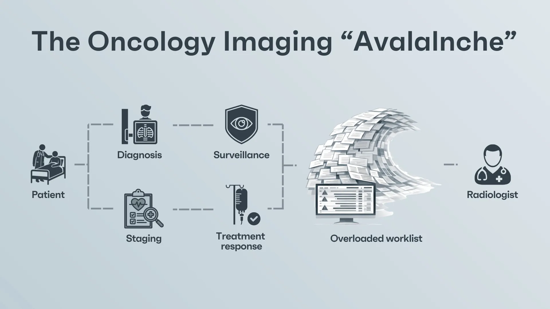
Why detection is the first step, but no longer enough
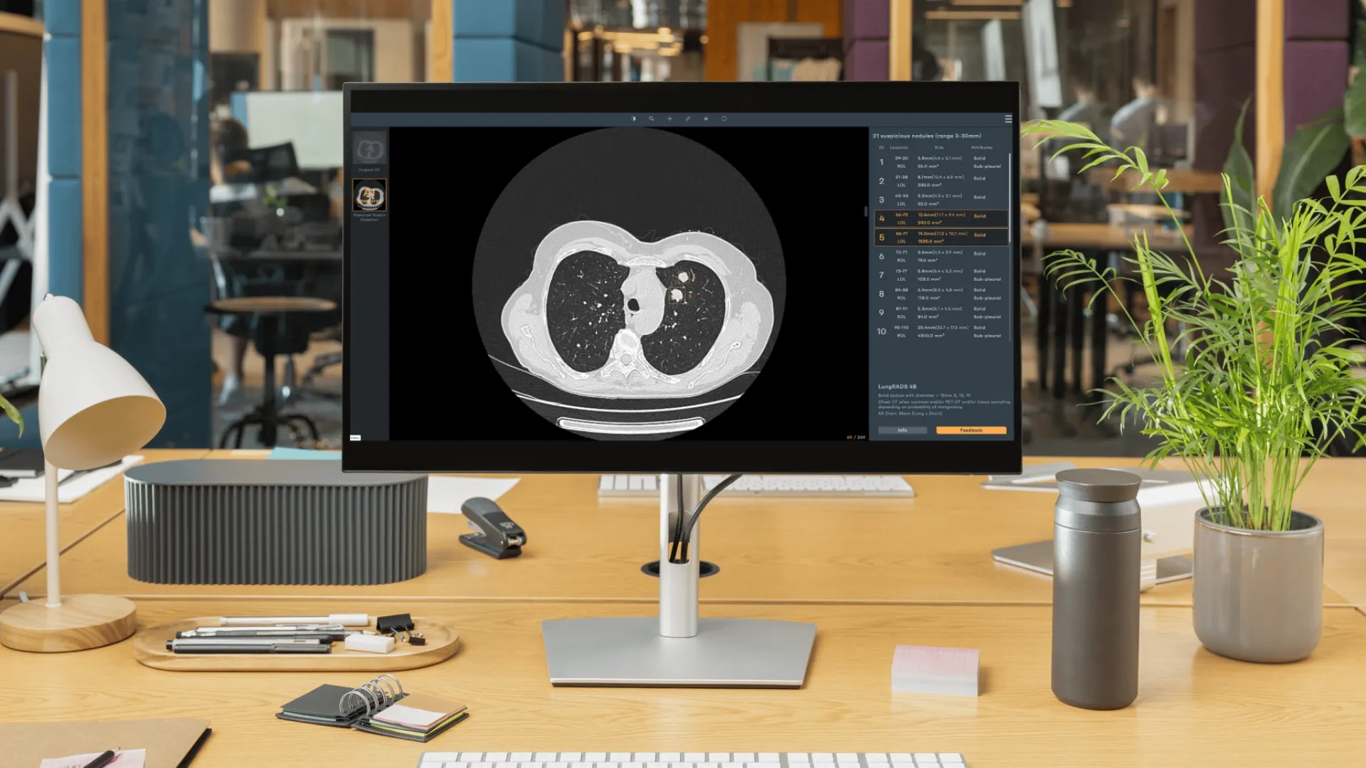
Current AI tools excel at pointing out super-specialised potential abnormalities (lung nodules, spine fractures, liver masses, etc), but they operate in isolation. They do not know whether a finding is new or pre‑existing, whether its growth rate is accelerating, whether it lies alone or belongs to the patient’s cancer type. Without context, a flagged finding can be clinically meaningless or even distracting. For example, a chest CT may contain dozens of benign pulmonary nodules; highlighting them all adds to the workload rather than reducing it. Conversely, a subtle metastasis growing within an adrenal gland on abdominal CT may be lost among numerous false positives if the system lacks an understanding of the patient’s disease.
Oncology care requires more than detection, as it demands prioritisation. A lung lesion that has doubled in volume since the last scan is more urgent than a stable scar. A new colon mass in a patient with familial cancer history may alter therapy, whereas another tiny cyst may not. The role of AI must therefore evolve from simple marking to tracking and ranking. Systems need to assign urgency scores to findings, reorder the worklist based on potential impact, and alert clinicians to changes that warrant immediate action. Prioritisation also helps filter out benign or irrelevant findings, allowing radiologists to focus on the cases that matter most.
From triage to contextual prioritisation
The next generation of AI CT scan analysis will incorporate sophisticated triage mechanisms. Rather than processing studies in the order they were acquired, algorithms will determine which cases need attention first. For instance, a follow‑up CT showing multiple new lesions in the liver and lungs should jump to the top of the queue, while a routine postoperative check might remain lower on the list because the AI didn't find anything suspect or urgent. The system could adapt its prioritisation dynamically as new studies arrive or as preliminary results are reviewed.
Contextual triage requires AI companies and their models to integrate information across different modalities, different types of studies (contrast and non-contrast / low-dose and normal dose CT) and multiple body regions, not just a single organ. Why? Because this is the way medical professionals evaluate. Thoracic, abdominal and pelvic findings often have interdependent significance. Enlarged mediastinal lymph nodes may suggest systemic spread from a pelvic tumour; bone metastases in the pelvis might indicate progression of a previously stable prostate cancer. An AI platform must therefore maintain a holistic view of the patient’s anatomy. This is particularly relevant in the era of total‑body imaging and whole‑body PET/CT, where a single acquisition spans from head to thigh. Even if detection across individual regions is solved, the system’s ability to connect the dots between them determines clinical value.
Multi‑lesion, multi‑organ context: beyond single nodules
Cancer rarely appears as a solitary lesion. Solid tumours metastasise to multiple sites; haematological cancers present with numerous foci. Treating these diseases means understanding both the aggregate tumour burden and the heterogeneity among lesions. A patient may have a dominant liver metastasis that responds to therapy while smaller lung nodules remain unchanged. Some lesions may harbour resistant clones and behave differently from others. Focusing solely on a single “target lesion” is insufficient.
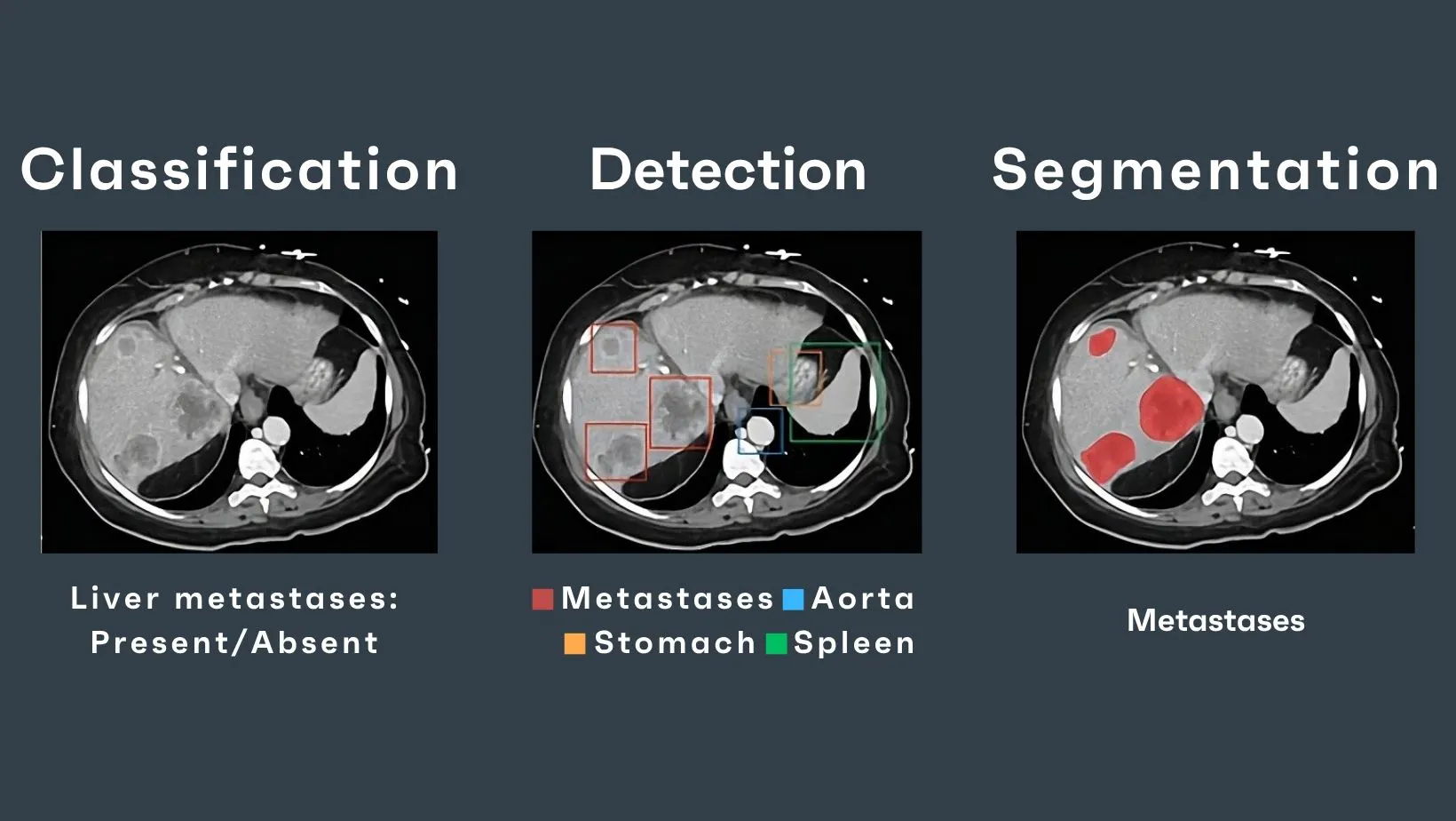
At Rayscape, we think modern AI systems must therefore quantify and interpret multiple lesions across organs. This involves accurate segmentation of each lesion, assignment of an anatomical label (e.g., lung, liver, bone), measurement of volume, characterisation of the lesion, and comparison with prior scans. Crucially, the system must recognise relationships among lesions: Are they part of the same disease process? Do they share imaging signatures? Are they responding similarly to treatment? Answering these questions can inform therapy decisions, such as continuing systemic therapy versus switching to local ablation.
Handling multi‑lesion context also includes managing the explosion of incidental findings. Screening programmes generate large numbers of benign or indeterminate nodules. Algorithms must learn to differentiate clinically significant lesions from inconsequential ones and adjust their alarms accordingly. Part of this involves training on diverse datasets that capture the variability of normal anatomy across the thorax, abdomen and pelvis. Incorporating multi-region database training will refine these assessments further.
Lesion tracking and longitudinal follow‑up
A core component of oncology imaging is longitudinal assessment: deciding whether tumours are shrinking, growing, or holding their ground. In practice, this means returning to the same patient again and again, the same anatomy, the same battleground, and asking what has changed since the last chapter. Traditionally, radiologists select a handful of target lesions, measure their diameters, and compare them manually across time. It works, but it is slow, reductive, and unfair to the full complexity of disease: countless lesions remain unmeasured, unnamed, and therefore almost invisible to the structured logic of follow-up. A three-dimensional tumour becomes a single line. Irregular growth becomes statistical noise. What does not fit the measurement framework risks becoming a footnote. AI changes the nature of follow-up by turning lesion tracking into an automated memory. Instead of relying on a few manually chosen targets, algorithms can map tumour burden across the entire scan volume, measuring lesions in three dimensions and tracking them through time with volumetric precision. Advanced registration aligns serial CT studies even when the patient’s positioning shifts, when body habitus changes, or when the imaging protocol evolves. By matching lesions across timepoints, AI can detect new metastases, quantify subtle changes in existing disease, and separate treatment-related transformation, necrosis, scarring, and inflammatory pseudoprogression from true progression.
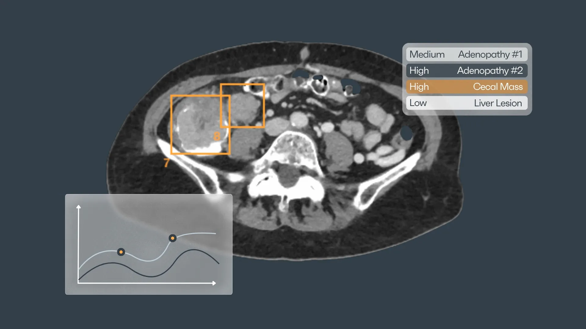
Beyond measurement, longitudinal models can learn temporal behaviour. When these patterns are modelled across multiple lesions rather than isolated targets, the follow-up scan becomes a trajectory. AI can forecast where the disease appears to be heading, highlight early relapse signals before they become obvious, and propose imaging intervals or treatment checkpoints that align with risk, all synchronised in real-time with international guidelines.
Integrated AI decision support is the backbone of Radiology
Detection, triage and tracking generate large amounts of structured data. The value of these data lies in their integration into a coherent clinical narrative. Best AI for radiology platforms must therefore include decision support that synthesises imaging findings with clinical records. Such systems can provide personalised risk assessments, recommend appropriate imaging follow‑up intervals and highlight potential complications.
An effective decision support module update for the basic AI software that is deployed in a hospital might, for instance, recognise that a new adrenal lesion in a patient with a known lung primary has a high likelihood of being metastatic and recommend biopsy or targeted therapy. It could flag a cluster of small hepatic lesions as suspicious in a patient with colorectal cancer, prompting an earlier oncology consultation.
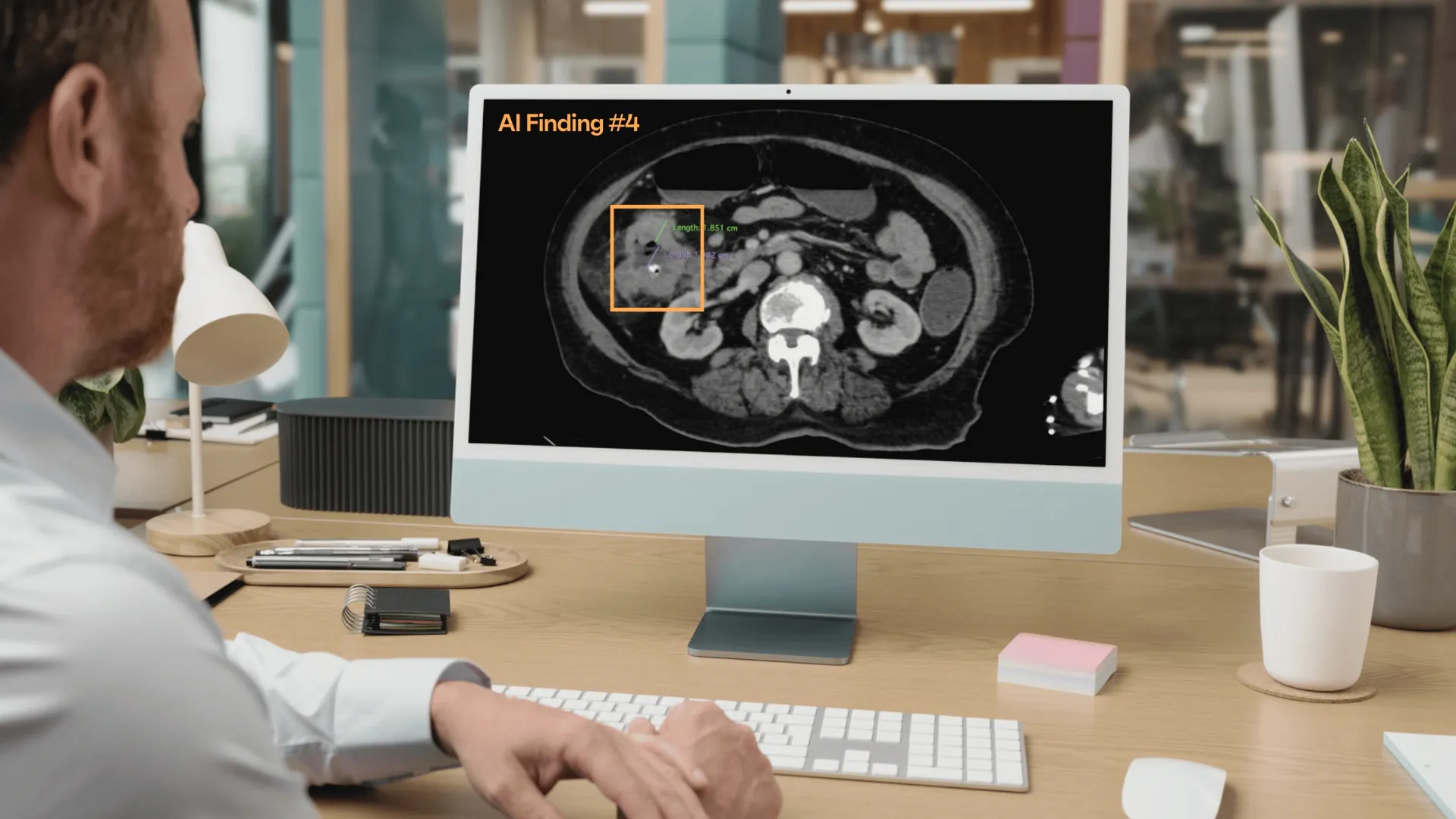
Importantly, decision support must be transparent and explainable. Radiologists need to understand why a system prioritised a case, flagged a lesion or recommended a management step.
Rayscape's blueprint for the next‑generation AI radiology platform
To solve the oncology and cancer screening worklist problem, an AI platform should incorporate the following architectural elements:
- Unified multi‑region detection: A single pipeline should analyse thoracic, abdominal and pelvic regions without requiring separate models. Shared feature representations enable cross‑region consistency and reduce computational overhead.
- Active worklist management: The platform should continuously monitor incoming studies, assign urgency scores based on imaging and clinical parameters, and reorder the worklist accordingly. Alerts for critical findings should be routed immediately to the responsible clinicians.
- Comprehensive segmentation and annotation: Accurate segmentation of organs and lesions is essential for volumetric assessment and anatomical localisation. The system should produce standardised labels that integrate seamlessly with reporting tools.
- Longitudinal registration and tracking: Robust algorithms must align serial scans, match lesions across time, and calculate volumetric and morphological changes. They should also handle differences in patient positioning and imaging protocols.
- Multi‑lesion analytics: Aggregating metrics across all lesions can provide insights into total tumour burden, heterogeneity and treatment response. Statistical models and machine learning should combine these metrics to predict outcomes and guide therapy adjustments.
- User‑centric interface: Radiologists and oncologists should be able to interact with the AI system through intuitive dashboards. Customisable views can highlight the most urgent cases, show temporal changes in tumour volume and provide context‑aware recommendations.
- Continuous learning: The system should learn from new data and user feedback. As oncology practice evolves, algorithms must adapt to emerging therapies and new imaging modalities while maintaining robustness and safety.
Conclusion
When detection algorithms have matured to the point where they no longer represent the main obstacle in radiology. The new challenges are rooted in scale and context: prioritising a deluge of scans, interpreting multiple lesions across the thorax, abdomen and pelvis, and delivering actionable insights that change patient management. Addressing the oncology worklist problem requires a holistic approach that combines detection with triage, tracking and decision support. AI radiology for cancer imaging will fulfil its promise only when it evolves from a collection of detectors to an integrated platform that understands patient journeys, guides clinical decisions and empowers physicians to deliver personalised care at scale.
Rayscape’s foundation in thoracic imaging has given us direct exposure to how artificial intelligence behaves inside real clinical environments. Chest CT became a proving ground for understanding radiologic complexity itself. In the lungs, we learned how models cope with motion, heterogeneous aeration, overlapping anatomical structures, and the constant challenge of separating true pathology from artefacts or benign variants. We studied how faint texture shifts can precede overt lesion formation, how acquisition parameters influence conspicuity, and how performance subtly changes across scanners, protocols, and patient populations.
Over time, one insight became unavoidable: AI becomes clinically meaningful not through isolated detections, but through consistent interpretation across thousands of heterogeneous studies. Radiologists don’t build trust around occasional brilliance; they build it around stability, reproducibility, and explainability. Rayscape CXR and LungCT gave us the clinical feedback loops needed to refine those principles. It showed us what it means for AI to operate in regions where normal variation is broad, early lesions are faint, and disease patterns rarely follow neat visual categories.
Expanding our AI into multiple regions is an extension of what radiologists already do: synthesising weak signals across organ systems, resolving competing findings, and tracking disease as a continuous process rather than a set of isolated observations.
For clinical teams exploring how advanced imaging intelligence can support prioritisation, lesion tracking, and workflow coherence across oncology TAP-CT, Rayscape is already building in this direction. If you’d like to see what is currently available today, or discuss where this is heading next, we’re always open to thoughtful conversations with radiologists and institutions pushing oncology imaging forward.


Discover how Rayscape works
integrate Rayscape AI into
your hospital's infrastructure
and improve both your
daily workflow and patients' experience.
