

Thoracic, Abdominal & Pelvic CT AI for Oncology Detection
The human eye is a marvel of biological engineering, but it operates within fixed constraints. When a radiologist reviews a CT scan, they process images sequentially, comparing each slice against an internal library built from years of training and thousands of cases. This perceptual framework, honed through experience, excels at recognizing familiar patterns but struggles at the margins, where early malignancy hides in noise, where subtle texture changes fall below the threshold of conscious detection, where the signal is present but perceptually invisible.
Artificial intelligence operates differently. It does not "see" in the way humans do. Instead, it quantifies. Where a radiologist perceives a region as "slightly heterogeneous," an AI model computes the exact distribution of Hounsfield Unit values across thousands of voxels, measures the statistical deviation from expected tissue behavior, and compares that signature against training data spanning tens of thousands of annotated lesions. It detects patterns that exist mathematically but remain imperceptible to human vision, not because radiologists lack skill, but because the human visual system was never designed to process medical imaging at this granular, statistical level. And understanding the difference between human perception and algorithmic pattern recognition is essential to understanding how AI can meaningfully contribute to oncologic imaging.
The perceptual threshold: where human vision ends and computation begins
Radiologists rely on visual perception alongside decision-making under uncertainty, making errors inevitable, especially under constrained conditions. The diagnostic process begins with detection, at the moment when a radiologist's eye catches something that warrants closer inspection. But with this come perceptual errors, particularly "failure to detect," which represent the most common interpretive error and cause of litigation in radiology. These failures are not about incompetence because they stem from the inherent limitations of biological vision when confronted with statistical subtlety. A three-Hounsfield-Unit attenuation difference between a tiny hepatic metastasis and surrounding parenchyma may be mathematically significant but visually imperceptible during rapid review of a 500-slice dataset. The lesion is present. The radiologist simply cannot see it, not without knowing exactly where to look.
AI methods excel at automatically recognizing complex patterns in imaging data and providing quantitative rather than qualitative assessments of radiographic characteristics. An AI model trained on thousands of hepatic lesions learns the statistical signature of early metastatic disease: the subtle clustering of attenuation values, the micro-textural patterns that distinguish true pathology from benign perfusion variation, the geometric deviations that suggest infiltration rather than normal anatomic variation. It flags regions that fall outside expected probability distributions, even when those deviations are too subtle for the human eye to consciously register. This is where AI becomes clinically valuable, not by replacing radiologic judgment but by expanding the perceptual bandwidth. It surfaces weak signals that might otherwise remain hidden in the shadows of normal variation.
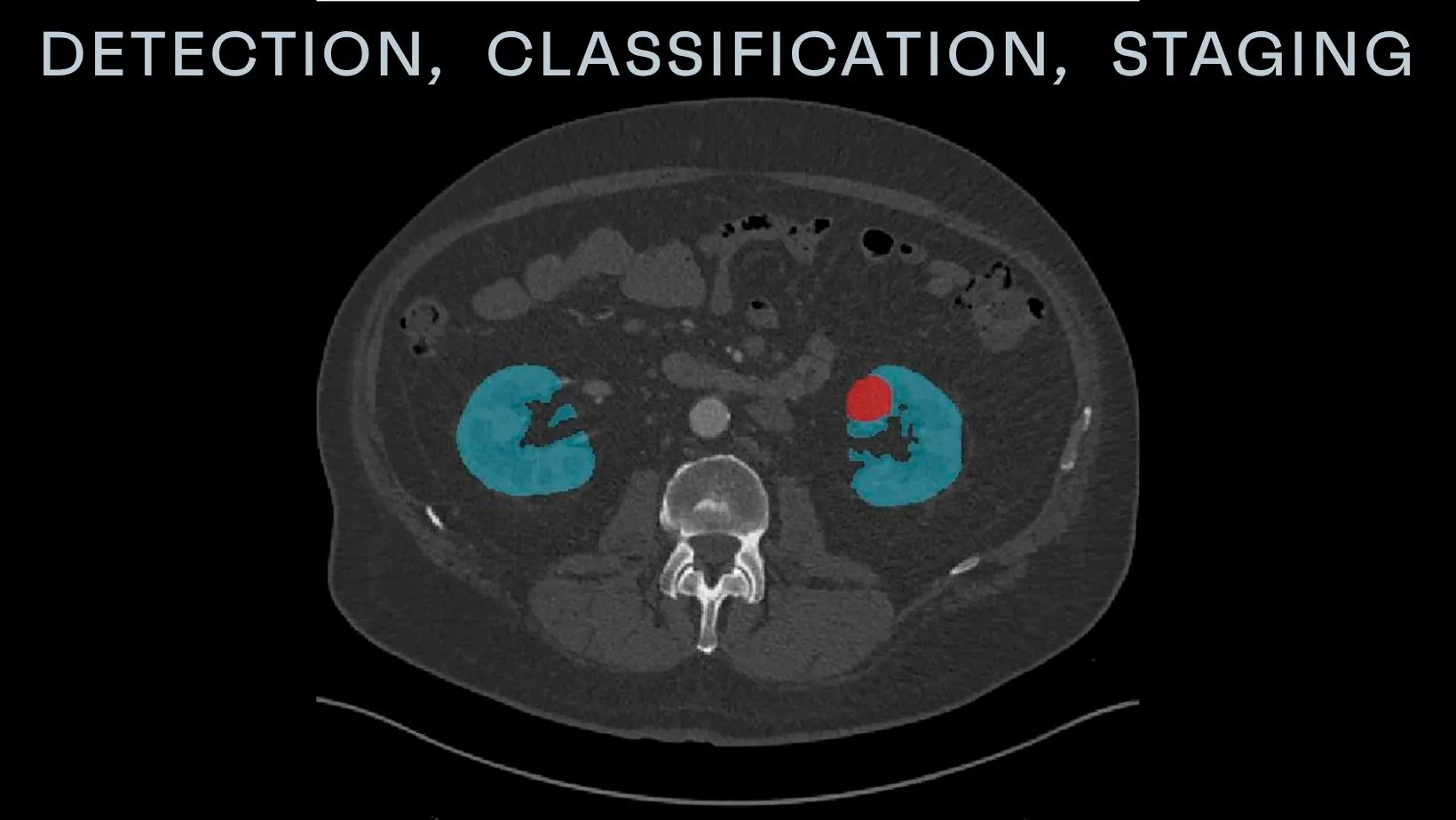
Subtle oncologic findings where AI consistently improves visibility
The clinical evidence for AI's impact on subtle lesion detection continues to accumulate across multiple organ systems and pathologies. In thoracic imaging, AI already improves diagnostic accuracy and reduces the risk of misdiagnosis by identifying subtle patterns and anomalies that may be missed by the human eye. But what the next step is, the real test, it comes in anatomically complex regions where density relationships shift continuously, and pathology often manifests as faint distortions rather than discrete nodules. Let's take pancreatic adenocarcinoma as an example, as its detection exemplifies this challenge. Early tumors frequently appear as vague hypoattenuating regions that blend into the gland's natural lobulated texture, visible only as secondary signs: ductal cutoff, upstream dilation, or loss of the characteristic enhancement pattern. AI trained on multi-phase pancreatic protocols learns to recognize these constellation findings across arterial, venous, and delayed phases, flagging cases where the statistical probability of malignancy exceeds a learned threshold, even when no definitive mass is visible.
Small hepatic metastases present a different challenge: phase-dependent conspicuity. Hypervascular metastases flash-enhance in the arterial phase but may become nearly isodense in the portal venous phase. Hypovascular lesions behave oppositely. A radiologist reviewing only the portal venous phase might miss an arterial-enhancing focus entirely. AI algorithms need to be trained on large ground truth datasets so they can autonomously detect and flag suspicious lesions, potentially allowing radiologists to accelerate review and improve productivity. By processing all phases simultaneously and computing enhancement kinetics for every voxel in the liver, the algorithm should identify lesions that might be dismissed as noise in any single phase but demonstrate statistically significant enhancement behavior when analyzed temporally.
The value proposition is straightforward: AI does not get tired. It does not experience perceptual saturation. And it maintains the same level of vigilance on the 500th slice as it did on the first.
Why interpretability and validation matter for clinical trust
Despite promising performance metrics, AI adoption in radiology remains limited. Surveys find only a few percent of clinics use AI tools routinely. The reason is not technological skepticism, but the absence of trust, and as we learned, trust in clinical AI is built on three foundations: interpretability, validation, and transparency. Interpretability in radiology is essential because wrong diagnoses can have severe consequences for patients, and clinical diagnosis is often not trivial and prone to interpretation errors. When an AI model flags a region as suspicious, the radiologist needs to understand why. Is the algorithm detecting a real pattern, or has it mistaken a benign variant, artifact, or normal structure for pathology? Without explainability, the radiologist is left in an uncomfortable position: trust the machine blindly or ignore it entirely. And neither approach is acceptable or easy to make.
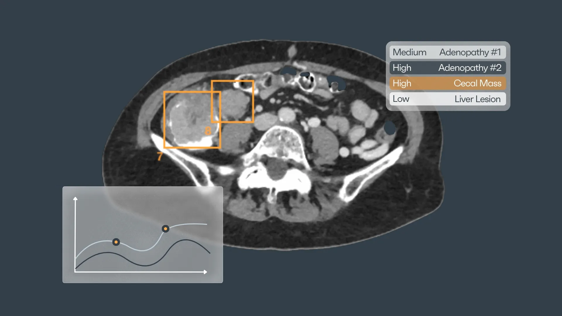
Modern interpretability methods address this through visual explanations, bounding boxes, heatmaps that highlight the specific image regions driving the algorithm's prediction, activation maps that show which learned features are triggering the detection, or confidence scores that quantify certainty, either being percentages or grades. Explainability enhances the diagnostic ability of radiologists because it offers them an informed interaction between a human and a computer.
But interpretability alone is insufficient. It would have been too easy, wouldn't it? Verified datasets are crucial to provide valuable support, yet algorithms trained on standardized datasets lack the ability to adapt to local knowledge. This is why validation matters, from internal validation on the same population where the model was trained, to external validation across diverse scanners, protocols, and patient demographics - a generalization opportunity. Some companies with AI systems trained on a single country's data may perform brilliantly locally, but fail catastrophically when deployed elsewhere, due to differences in scanner calibration, contrast protocols, patient populations, or imaging techniques.
What we learned from thoracic AI: accuracy, adoption, and workflow integration
Rayscape's foundation in thoracic imaging provided years of practical insight into how AI behaves in real clinical environments. Lung nodule detection was an ideal starting domain: relatively well-defined pathology, clear density contrast against surrounding air, and decades of clinical research establishing ground truth. But even in this favorable environment, developing clinically useful AI required confronting realities that pure performance metrics do not capture.
Accuracy is necessary but not sufficient. Early in our development at Rayscape, we learned that high sensitivity and specificity on benchmark datasets did not guarantee clinical value. If the algorithm detected nodules that radiologists already identified easily, it added no value. If it flagged every calcified granuloma, fat island, and vascular cross-section as suspicious, it generated workflow friction rather than assistance. The real challenge is calibrating the model to flag only the findings that warranted radiologist attention—nodules that were genuinely subtle, borderline in size, or questionable in morphology.
This required iterative refinement with radiologist feedback. We did not simply train a model and deploy it. We embedded it in live workflow, collected data on which detections radiologists found helpful versus distracting, and continuously adjusted sensitivity thresholds and false positive suppression mechanisms. The goal was not perfect sensitivity, because in reality, no algorithm achieves that, neither the radiologists, but optimal clinical utility: maximizing actionable detections while minimizing noise.
A technically brilliant algorithm that requires radiologists to open a separate application, manually load cases, and review results outside their normal reading environment will not be used in daily practice. The radiologist doesn't have either the time or the patience for this. We learned this through early pilot implementations where adoption rates plateaued despite radiologist enthusiasm for the technology. The problem was friction, as even small amounts of additional effort created enough resistance to derail habitual use.
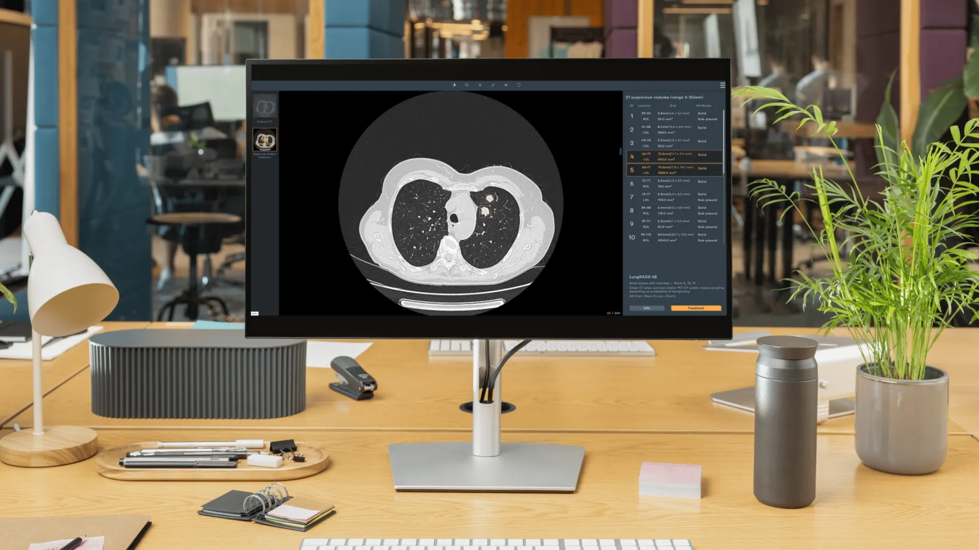
The solution was seamless PACS integration. The AI needs to operate invisibly in the background, processing cases automatically as they arrive, and presenting findings directly within the radiologist's existing workspace. Detections that appear as annotated overlays on the images themselves, with measurements, volumetric data, and longitudinal tracking integrated into the report workflow. Radiologists do not need to think about "using the AI", but see it as a simple part of the environment, like window/level controls or multiplanar reconstruction, something that feels natural.
Longitudinal analysis transforms screening workflows
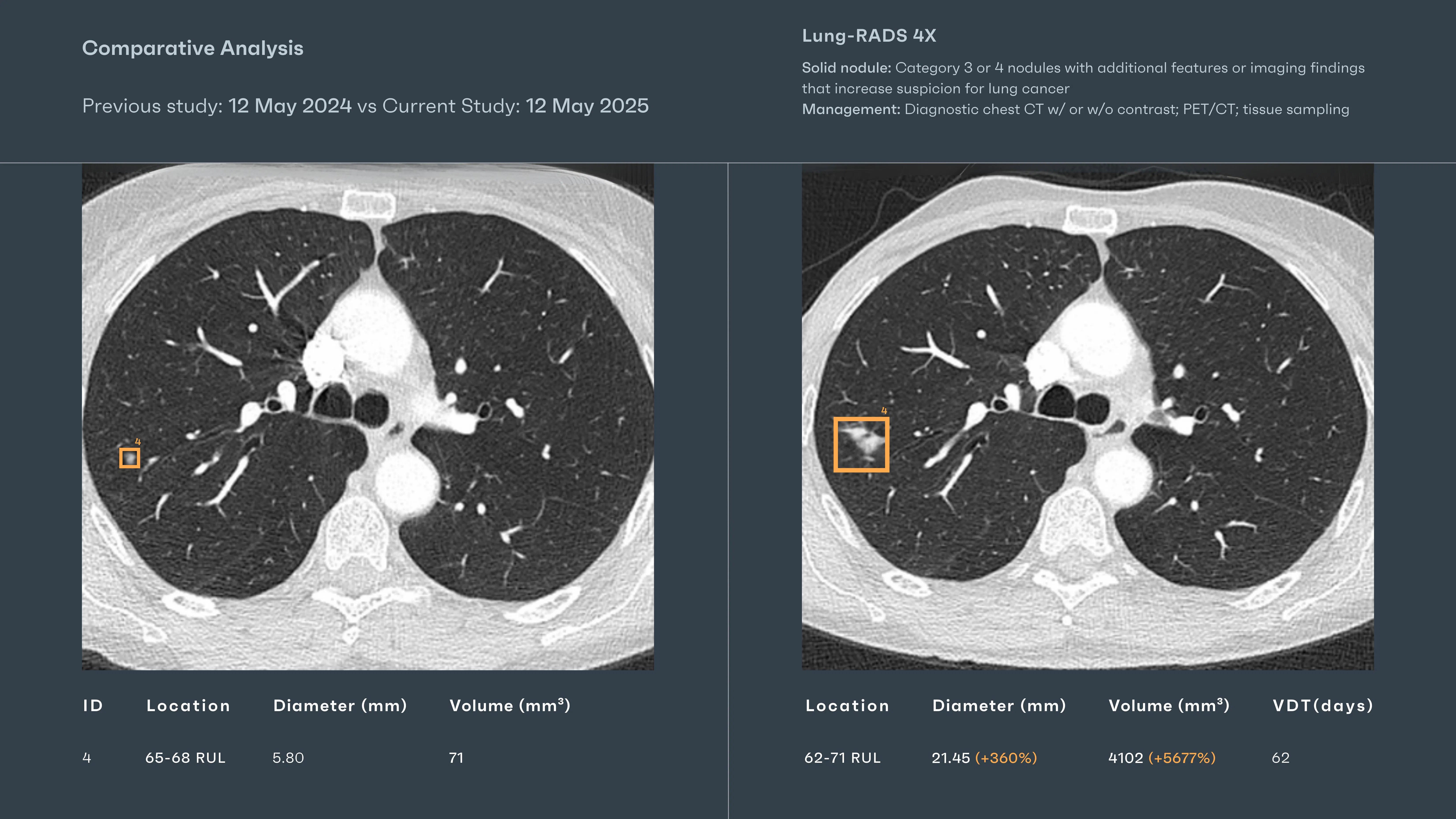
One of the most valuable capabilities we developed was automated nodule tracking across serial exams. In lung cancer screening, the critical question is not simply "Is there a nodule?" but "Has this nodule grown?" Manual measurement across multiple time points is time-consuming and subject to inter-observer variability. AI-powered volumetric tracking removes this burden: the system automatically identifies corresponding nodules across exams, quantifies volumetric growth, and displays side-by-side comparisons with precise measurements.
This capability proved transformative for screening programs managing thousands of follow-up scans. Radiologists could instantly assess whether a nodule met growth criteria for biopsy or remained stable, without hunting through prior reports or manually measuring multiple lesions across different exams. The workflow time savings were substantial, but the real value was consistency—eliminating the cognitive bias and measurement variability that plague manual tracking. Thoracic imaging follows well-established guidelines: Lung-RADS for screening nodules, Fleischner criteria for incidental findings. We integrated these classification systems directly into the AI output, so radiologists received not just detections and measurements but also guideline-based recommendations.
Extending to Abdominal and Pelvic Imaging: same principles, new challenges
The computational strategies that proved effective in thoracic imaging—volumetric modeling, multi-phase reasoning, subtle attenuation analysis, and longitudinal tracking—translate directly to abdominal and pelvic oncology. The underlying challenge is identical: detecting malignancy at its earliest, most treatable stage by identifying statistical patterns that fall below the threshold of reliable human perception. But the complexity escalates dramatically. Thoracic datasets have favorable characteristics: clear density stratification between air, soft tissue, and bone; relatively limited motion; and pathology that often presents as discrete nodules with definable margins. Abdominal imaging presents none of these advantages.
Multiple organs with overlapping densities coexist in proximity. Enhancement kinetics shift continuously across arterial, venous, and delayed phases. Motion from respiration, vascular pulsation, and bowel peristalsis introduces artifacts and blur. Partial volume effects at organ interfaces mimic pathology. Normal anatomic variants in one region resemble malignancy in another. Fat planes that should separate structures are frequently obliterated by inflammation, prior surgery, or desmoplastic reaction.
The dataset requirements alone exceed what thoracic imaging demanded. Training a model to distinguish early pancreatic adenocarcinoma from age-related fat infiltration and normal parenchymal heterogeneity requires tens of thousands of annotated pancreatic CT exams, spanning multiple scanners, protocols, and patient demographics. The model must learn not just what pancreatic cancer looks like, but what normal pancreatic tissue looks like across a wide range of appearances—and how to distinguish malignant deviation from benign variation.
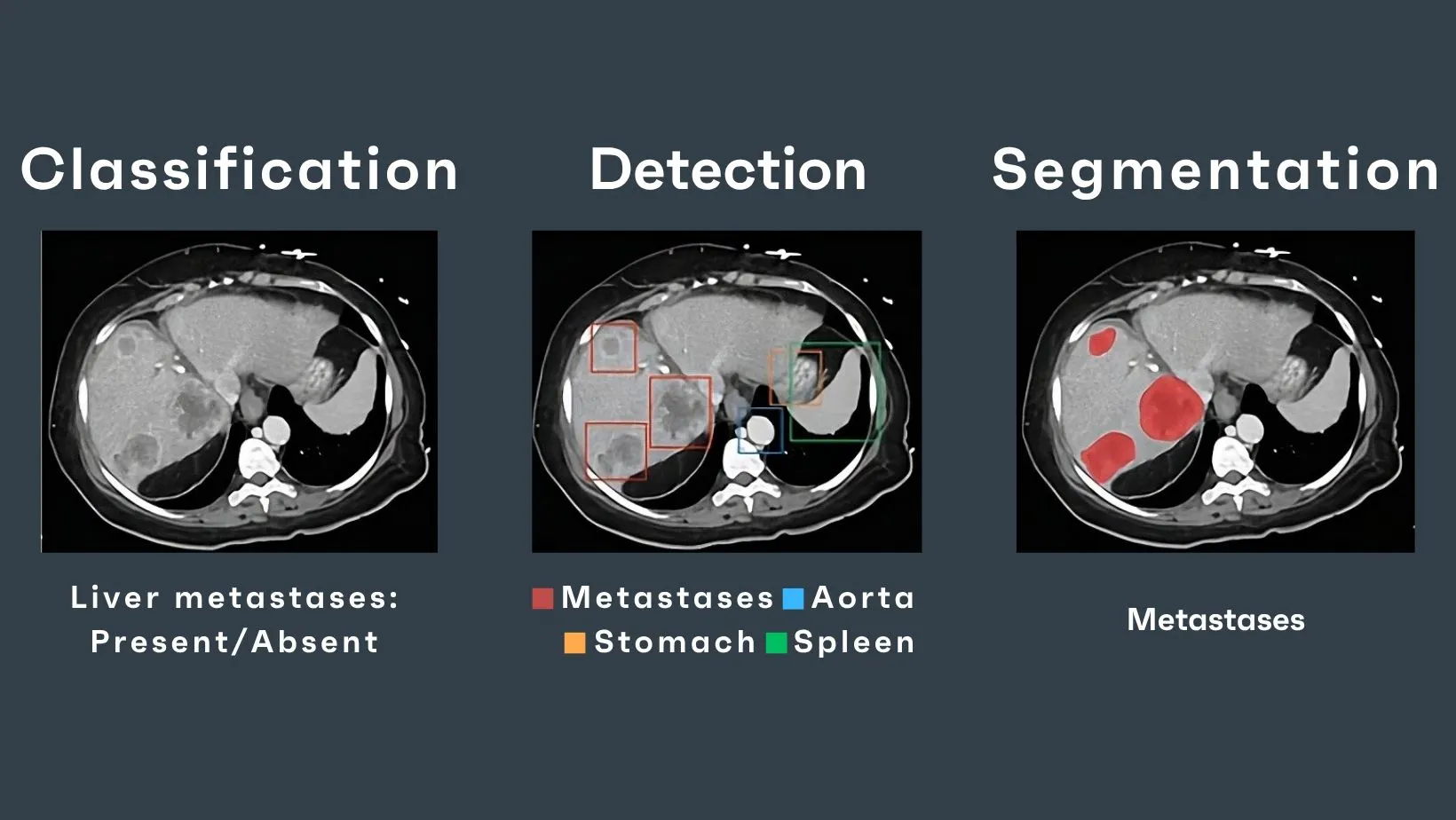
Liver metastasis detection presents a different challenge: the same lesion may appear as a hyperdense flash-filling focus in the arterial phase, a subtle hypoattenuation in the portal venous phase, and nearly isodense in the delayed phase. The AI must integrate information across all phases, computing enhancement curves for suspicious regions and comparing those curves against learned signatures of metastatic disease. This requires not just spatial pattern recognition but temporal reasoning—analyzing how tissue behavior evolves across multiple imaging time points.
These are solvable problems, but they demand far richer training data, more nuanced annotation, and more sophisticated model architectures than thoracic imaging requires. The path forward is clear, but it is also laborious, requiring extensive clinical collaboration, iterative refinement, and rigorous validation across diverse datasets.
Beyond the Lungs: Rayscape's Oncology Direction
For the past several years, Rayscape CXR and LungCT has been deployed in clinical environments, processing hundreds of thousands of thoracic scans and refining our understanding of how AI integrates into radiology workflows. That foundational model has taught us what matters: not just algorithmic performance on benchmarks, but reliability in production, interpretability in clinical practice, and seamless integration into existing workflows.
We are now extending those principles beyond thoracic imaging into multi-organ, multi-region oncologic detection. The focus is full thoracic, abdominal, and pelvic CT, where malignancy often hides within anatomical complexity and diagnostic challenges routinely surpass thoracic imaging in subtlety and heterogeneity. This is what we think is a natural progression. Oncologic imaging does not respect anatomical boundaries. Disease spreads along vascular pathways, lymphatic chains, and peritoneal surfaces. A comprehensive AI system for oncologic detection that follows the same logic radiologists use: synthesizing information across organ systems, integrating subtle findings from multiple regions, and tracking disease as a continuum rather than a collection of isolated detections.
At RSNA2025, we have offered an early look at this direction: multi-organ, multi-region lesion AI (artificial intelligence) detection for entire thoracic, abdominal, and pelvic oncology, a glimpse of where our research is heading, and the capabilities we are building toward.
The results are encouraging. A model that demonstrates sensitivity to subtle hepatic lesions across multiple enhancement phases, detection of small pancreatic masses that radiologists frequently revisit in retrospective review, and identification of peritoneal implants that standard reading workflows commonly miss is something that we think radiologists would find useful in their daily workflow. By demonstrating that the same principles that made LungCT clinically valuable—volumetric reasoning, multi-phase integration, and workflow-embedded detection- can extend to anatomically dense and diagnostically subtle regions.
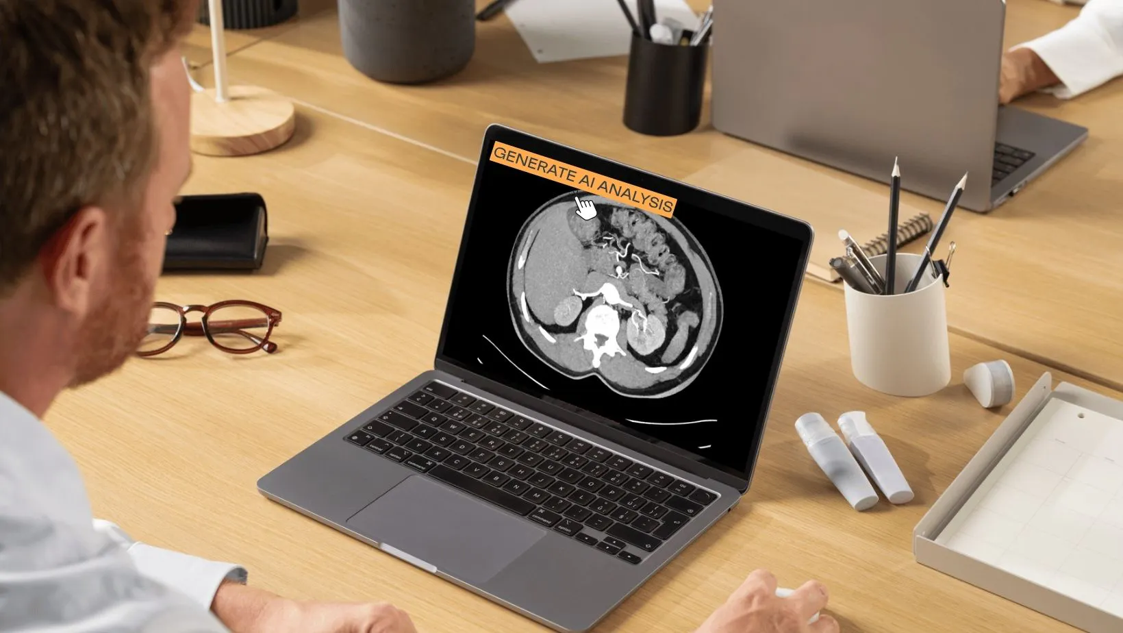
This development direction reflects a simple reality: the future of oncologic AI is not single-organ, single-pathology detection. It is a comprehensive, multi-region analysis that mirrors how radiologists actually read scans—synthesizing findings across anatomical compartments, integrating temporal data, and providing decision support that fits naturally into existing workflow patterns.
For institutions and clinical teams interested in how advanced AI can support their oncologic imaging workflows, we welcome conversations. Rayscape has always developed our technology in close collaboration with practicing radiologists, and that approach will define this next phase of work. If you are exploring AI solutions for abdominal and pelvic oncology, or if you are interested in learning more about our current and future development roadmap, we would be happy to discuss where our work intersects with your clinical needs.
The path from shadows to signals is long, but it is illuminated by collaboration between those who understand the clinical problem and those building the computational tools to address it. We are committed to walking that path thoughtfully, rigorously, and always with clinical utility as the guiding principle.


Discover how Rayscape works
integrate Rayscape AI into
your hospital's infrastructure
and improve both your
daily workflow and patients' experience.
