

Artificial Intelligence for lung Computed Tomography
Radiologists are medical doctors that specialize in diagnosing and treating injuries and diseases using medical imaging (radiology) procedures (exams/tests) such as X-rays, computed tomography (CT), magnetic resonance imaging (MRI), nuclear medicine, positron emission tomography (PET), and ultrasound. They get to see every interesting patient in the hospital from every single specialty, help make the diagnosis fast and move on to the next patient. In today’s blog post, we’ll continue the pulmonary medical imaging lesson that a radiologist learns for doing a chest computed tomography on a patient who may be experiencing lung issues.
CT is still the most commonly utilized and reviewed cross-sectional scanning technology for the thorax, and it is continuously evolving. Thoracic CT imaging techniques are adjusted to the clinical question and the structures of interest, much like any other imaging procedure.
AI for pulmonary analysis
To obtain images undistorted by respiratory motion artifacts, thoracic CT needs to be performed in a single breath-hold. The evaluation of a thoracic CT image dataset is no different from chest radiography in that more or less the same parameters and structures need to be evaluated. However, the emphasis, level of detail, and sequence of evaluation are tailored to the specific clinical question and are somewhat influenced by the CT acquisition that has been performed.
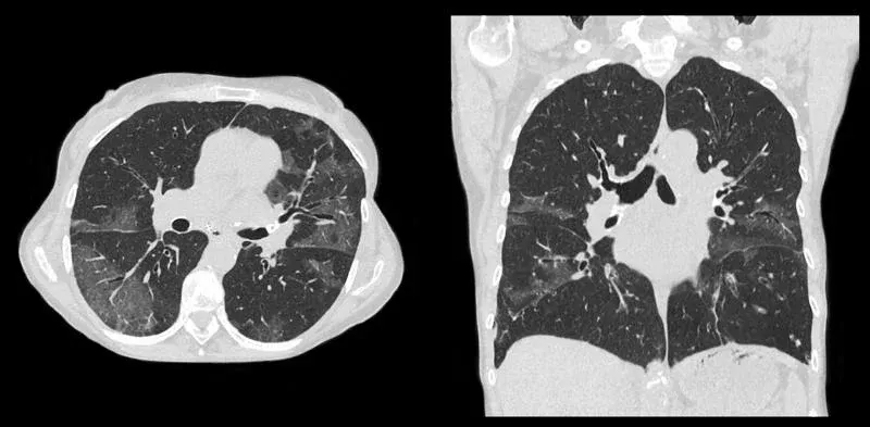
First, a doctor should always double-check that all of the examination’s essential datasets have been recreated and delivered to the PACS (either local or in cloud). They need to make sure that soft-tissue and lung reconstructions, thin and thick slice datasets, and coronal and sagittal reconstructions are available for review, and that the images are of sufficient diagnostic quality and are only minimally affected by respiratory motion and unwanted image noise.
If these characteristics are met then the radiologist can go further to interpret the image.
On CT, the placements of instruments such as endotracheal intubation and pleural drainage will be more precisely localized than on the X-ray. Artificial valves, aortic root implants, previous lobectomy, or surgical wedge resection can all be seen as evidence in the case of previous cardiothoracic surgery.
How to read a lung CT
The proximal airways will be first examined through lung sections to check that all lobar and segmental bronchi are intact and if there are any anatomical differences. It’s also vital to look for any endoluminal obstructive abnormalities in the airway space, particularly around bronchial bifurcations, where common cancers can be found. Proximal airway thickness, calcification, or nodularity is checked in case it could indicate tracheobronchial amyloid disease or other diseases caused by deposits.
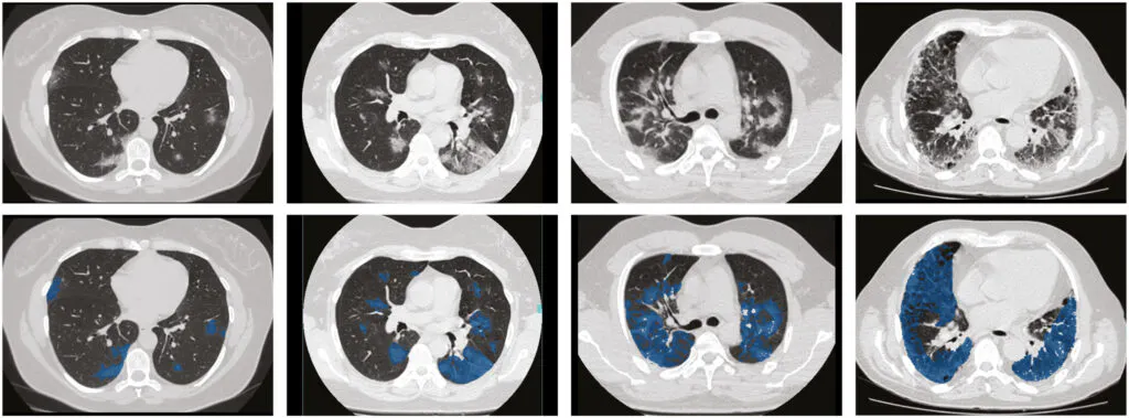
Finally, the distant airway diameter is measured to the adjacent pulmonary artery. Bronchitis is defined as a thickening of the airway wall relative to the arteries. If combined inspiratory and expiratory scanning is available, it is helpful to compare these images next to each other at this point to assess any dynamic airway changes that may indicate specific pathologies.
The first step in evaluating the lungs is to assess the volumes, noting any asymmetry in size. If a collapse is identified, it’s a good idea to check the airways that supply the area to make sure there’s no obvious obstructive condition. The pulmonary densities can then be examined to see if they are consistent and appropriate, taking into account the patient’s height, weight, level of inspiration, and intravenous contrast. Mosaic attenuation, or extreme heterogeneity in lung density, can be caused by any one or a combination of air trapping, interstitial lung disease, or pulmonary vascular disease.
Abnormalities on lung CT
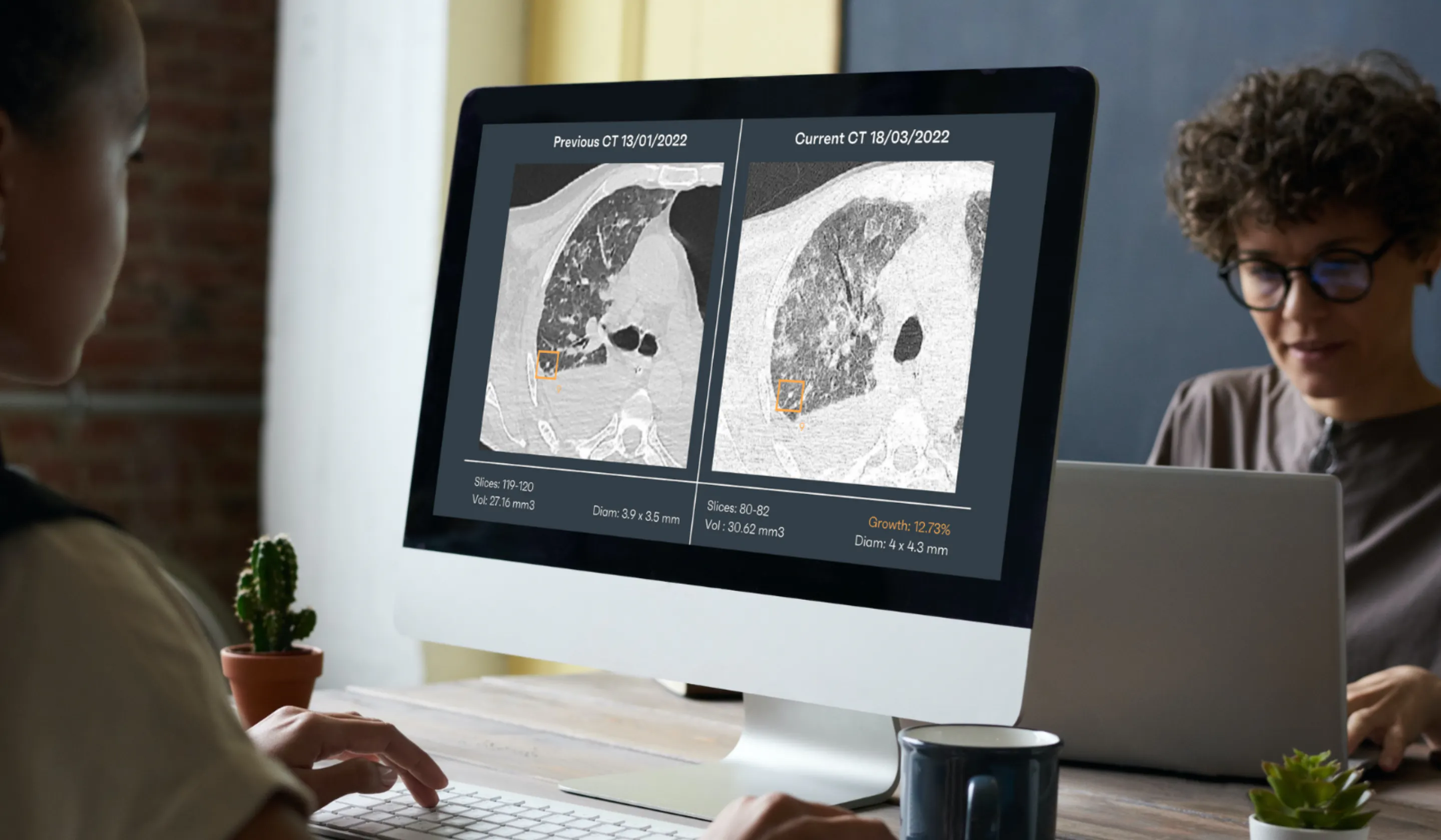
Various post-processing and image editing tools can be applied for certain scenarios when doing lung tissue evaluation. Maximum intensity projections, for example, enhance the search for pulmonary nodules, while volumetric analyses of pulmonary nodules, as well as prediction algorithms for the likelihood of malignancy, are progressively being integrated into protocols for follow-ups.
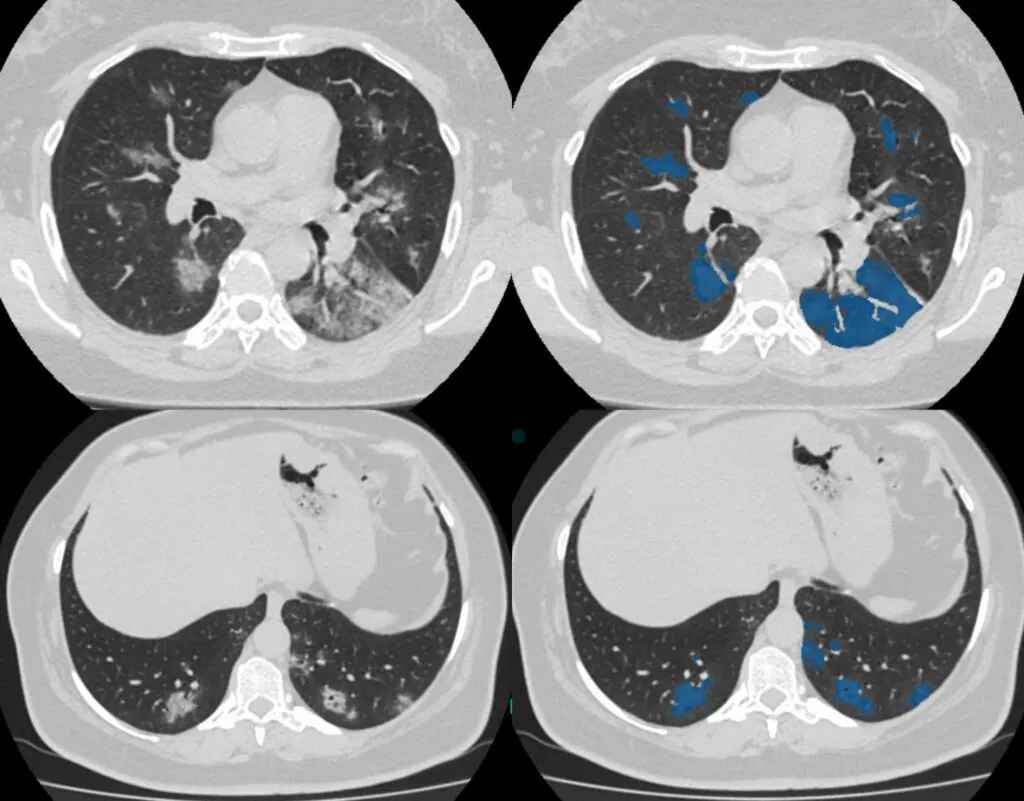
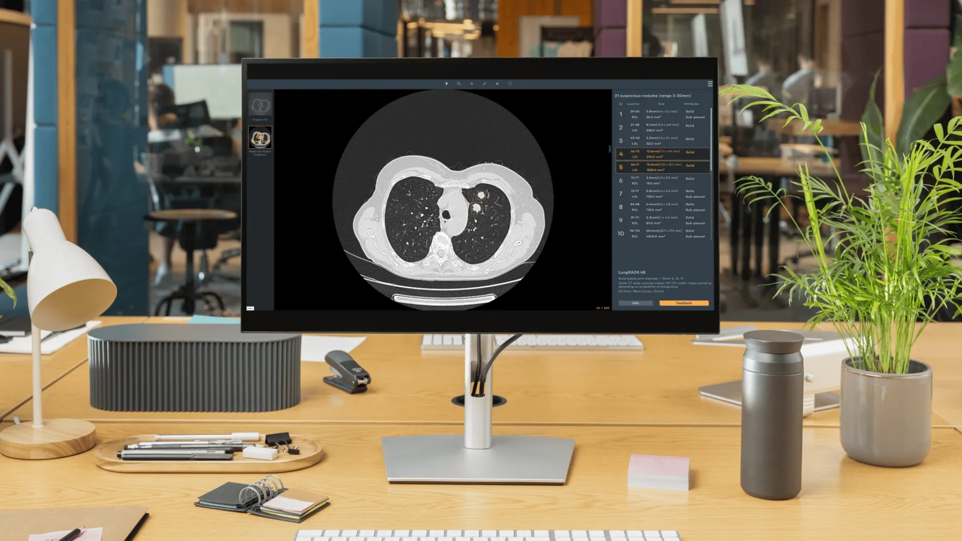
Rayscape provides a CT Nodule Detection algorithm that helps radiologists identify lung nodules that have a diameter between 3-30 mm. The algorithms highlight the presence of nodules on each slice and measure the diameter and volume of the identified ones separately.
The radiologist should then evaluate the median region of the chest. The anterior mediastinum could show remnant thymus material in teenagers or persistent proliferation in elderly people who have recently been sick or stopped taking corticosteroids. Thymus-derived cancers such as thymoma or lymphoma are examples of abnormal structures that could be seen.
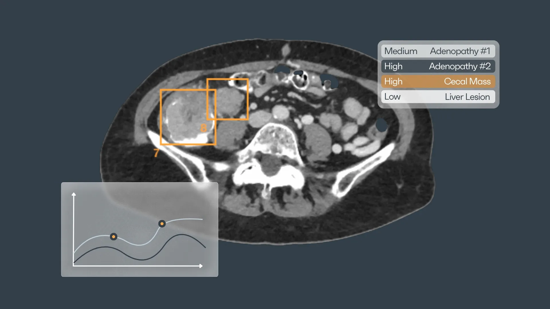
Any calcium deposit or growth of the mediastinal and hilar nodules is documented. Lymph nodes that measure 1 cm or above over a normal CT scale are usually regarded as enlarged. The location of any lymphadenopathy is evaluated, as the occurrence of bilateral nodule growth is associated with sarcoidosis.
Radiology tips for better results
Any nodal abnormality in other areas, such as the internal mammary and axillary territories, should be carefully examined because this could indicate undiagnosed breast cancer.
The evaluation of the chest circulation on a non-contrast CT is mainly limited to a qualitative judgment of how much the thoracic aorta (particularly the ascending aorta) is visibly dilated or highly calcified. Enlargement of the pulmonary arteries could also indicate pulmonary hypertension. A relevant metric for this is the ratio of the diameter of the pulmonary artery to the diameter of the ascending aorta.
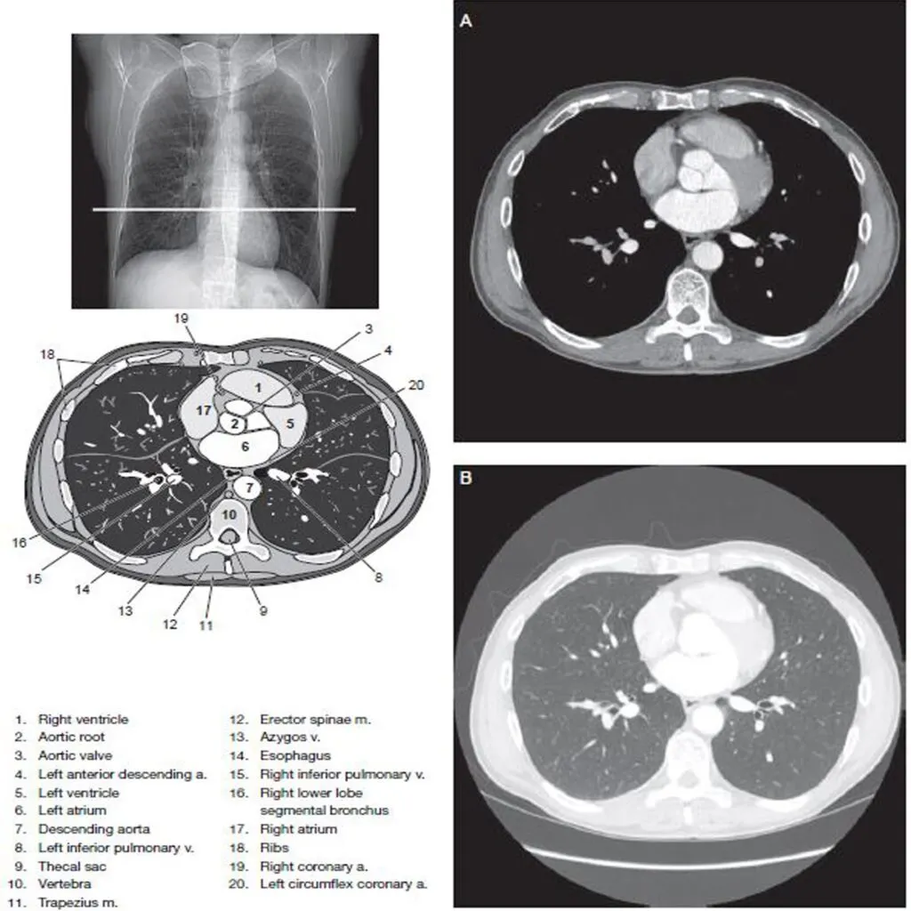
On contrast-enhanced CT, the aorta may be assessed for evidence of an acute aortic syndrome (dissection or penetrating atherosclerotic ulcer) and the pulmonary arteries for embolism.
Although a radiologist does not examine the heart in detail for a CT scan of the lungs, a quick look can occasionally reveal undetected intracardiac lesions or thrombi.
Non-contrast CT can easily detect pleural effusions, swelling, lesions, and nodules, either calcified or not. Pleural thickness, for instance, is particularly worrisome for a cancerous development. Inflammatory or malignant pleural diseases can benefit from delayed intravenous contrast (typically 60 seconds after administration).
A little quantity of pericardial liquid is typical in healthy people, but bigger fluid overload, especially when coupled with pleural, pulmonary, and airway defects, can indicate a systemic connective tissue condition like rheumatoid arthritis. Fluid density on CT is not a strong predictor of the origin, however, higher attenuation values of pleural and pericardial fluid may indicate an abscess or the existence of blood.
It’s important to check the diaphragm for every elevation which might signal a phrenic nerve paralysis, especially if there’s been trauma or if there’s a mass pinching or invading it. Similarly, the outline of each hemidiaphragm must be examined for any anomalies that could indicate a hernia.
It’s often a smart idea to examine the viewable abdomen for anomalies like retroperitoneal swollen lymph nodes or masses in the solid organs on the thoracic CT’s lowest scans.
In the end, each bone structure must be thoroughly examined.
Rayscape AI helps radiologists
As we can see, the detailed analysis of a chest CT scan involves many stages. Some of the steps mentioned and described above can be omitted, especially if the radiologist is at the end of a day’s work and fatigue is present.
Rayscape was designed to help radiologists be more, to be better. Our suite of products dedicated to the health of the lungs is seamlessly integrated into the doctor’s and hospital’s workflow, helping them to analyze chest x-rays and lung CTs faster and more efficiently.
Rayscape is an extra brain that allows you to heighten your ability to analyze. Book a demo today to learn about our solutions.


Discover how Rayscape works
integrate Rayscape AI into
your hospital's infrastructure
and improve both your
daily workflow and patients' experience.
