

COVID-19 Survivors Have Lung Lesions Two Years After Infection
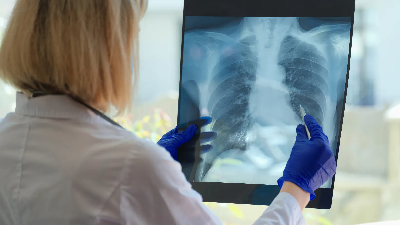
With over 600 million people having recovered from COVID-19 globally, concerns remain that some organs may suffer long-term damage, especially the lungs. Studies show that residual lung abnormalities may persist after hospital discharge, and these survivors should be followed up to detect and manage the pulmonary sequelae and functional impairment. Researchers in China conducted a prospective study to assess residual lung abnormalities and changes in pulmonary function at 6-month, 12-month, and 2-year follow-up periods.
Study findings: Residual lung abnormalities and changes in pulmonary function in COVID-19 survivors
The study population included 1251 COVID-19 patients who were consecutively discharged from Wuhan Jinyintan hospital between January 20th and March 10th, 2020. Ultimately, 144 participants were enrolled in the longitudinal study after applying exclusion criteria. Each participant underwent a chest high-resolution CT scan and completed a self-reported respiratory symptom questionnaire during each visit. Participants who completed lung function tests at three-time points were 110 at 6 months, 103 at 12 months, and 129 at 2 years. Pulmonary diffusion was considered abnormal when the diffusing capacity of the lung for carbon monoxide (DLco) was less than 75% of the predicted value.
Images were obtained using an unenhanced chest CT scan while participants held their breath immediately following full inhalation, and scans were performed from the level of the upper thoracic inlet to the inferior level of the costophrenic angle. The predominant CT pattern was categorized as ground-glass opacities (GGOs), consolidation, or reticulation predominant. The CT findings indicative of interstitial lung abnormalities (ILAs) included GGOs or reticular abnormalities, lung distortion, traction bronchiectasis, honeycombing, and non-emphysematous pulmonary cysts. The fibrotic ILAs were characterized by the presence of architectural distortion with traction bronchiectasis and/or honeycombing, whereas the non-fibrotic ILAs were defined as GGOs or reticular abnormalities.
The medical records of each participant in the acute phase of SARS-COV-2 infections were reviewed by one of three physicians. The study cohort included 62 participants from a previous study looking at chest CT findings upon hospital admission and at 6-month follow-up. The chest CT scans for each participant were performed at admission. During each follow-up, all participants underwent chest high-resolution CT scans and completed a self-reported respiratory symptom questionnaire. The pulmonary diffusion test was conducted on 110 participants at 6 months, 103 participants at 12 months, and 129 participants at 2 years.
The data were analyzed using SAS software. Quantitative variables were presented as mean ± standard deviation, and qualitative variables were expressed as frequency and percentages. The correlation between residual lung abnormalities and changes in pulmonary function was evaluated using the Spearman rank correlation coefficient. A p-value of less than 0.05 was considered statistically significant.
The importance of early detection and treatment of residual lung abnormalities in COVID-19 survivors
The researchers found that residual lung abnormalities after discharge from the hospital included ground-glass opacities, subpleural reticulations, cystic changes, traction bronchiectasis, honeycombing, parenchymal bands, and/or architectural distortion. These features fit the imaging definition of interstitial lung abnormalities (ILAs), which are potentially compatible with interstitial lung disease. The CT findings indicative of ILAs included ground-glass opacities or reticular abnormalities, lung distortion, traction bronchiectasis, honeycombing, and non-emphysematous pulmonary cysts. The fibrotic ILAs were characterized by the presence of architectural distortion with traction bronchiectasis and/or honeycombing, whereas the non-fibrotic ILAs were defined as ground-glass opacities or reticular abnormalities.
The study found that GGO and fibrotic-like change were found in 16.0% and 31.2% of COVID-19 patients, respectively, after 2 years of recovery. Furthermore, the study showed that a high percentage of participants had abnormal pulmonary diffusion function, indicating that the lung's ability to exchange gas was impaired. These abnormalities were more likely to occur in participants with residual lung abnormalities, especially in those with subpleural fibrotic ILAs.
This suggests that residual lung abnormalities may persist up to two years after COVID-19 infection. Clinicians should be vigilant in observing and managing the pulmonary sequelae and functional impairment in COVID-19 survivors, especially those with residual lung abnormalities. Further studies are needed to determine the long-term outcomes of COVID-19 survivors and to develop appropriate interventions. In the meantime, individuals recovering from COVID-19 should seek appropriate medical attention to prevent long-term complications.
Managing long-term sequelae in COVID-19 survivors: The role of artificial intelligence (AI) radiology software
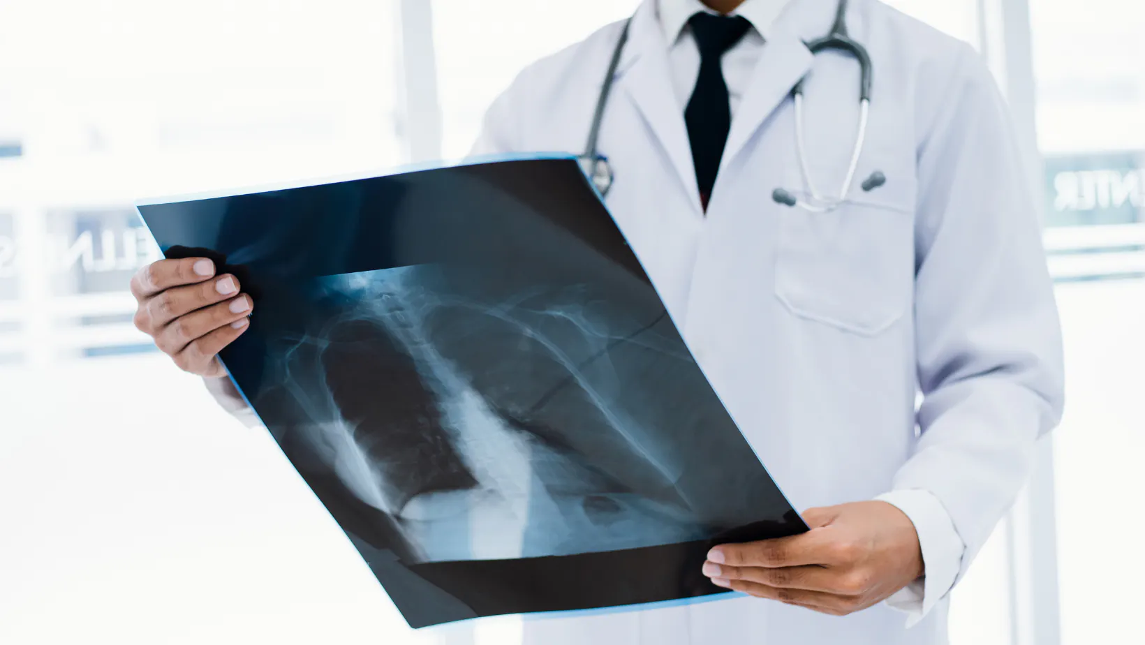
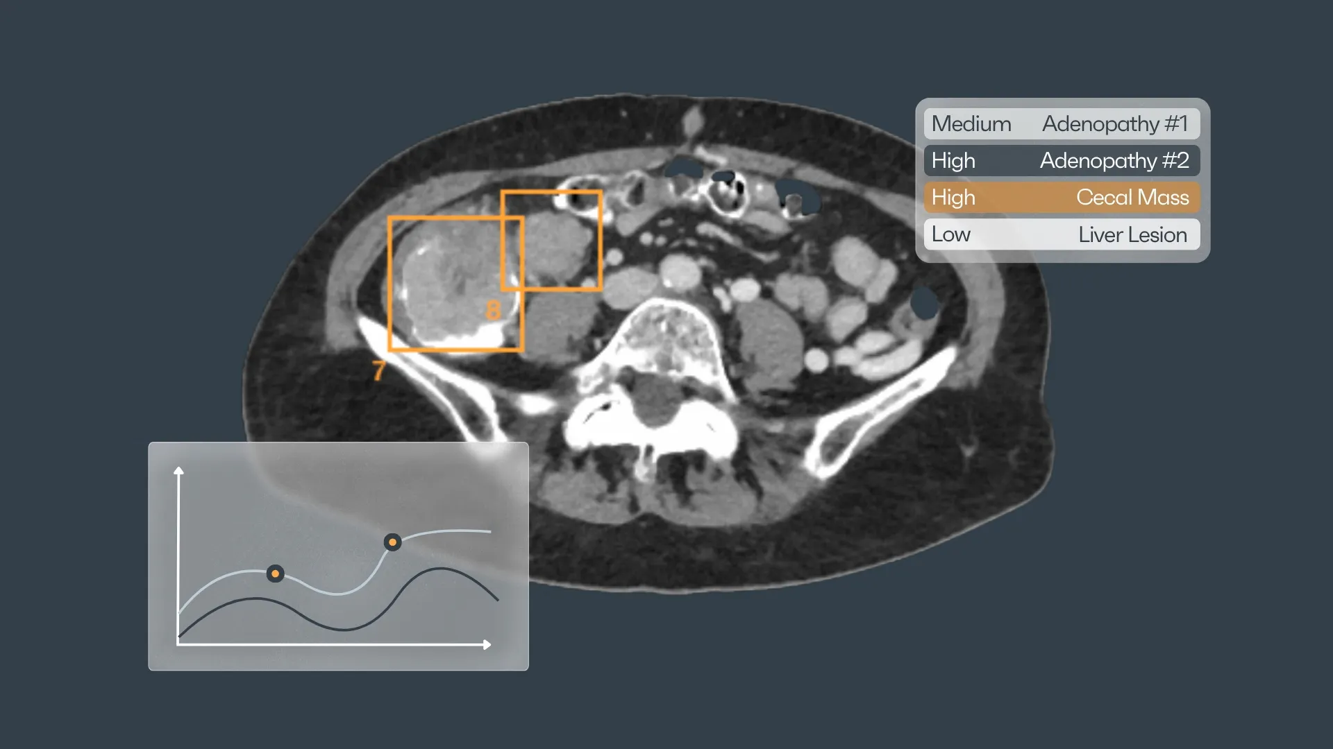
The study provides valuable insights into the residual lung abnormalities and changes in pulmonary function among COVID-19 survivors. These findings highlight the need for vigilant observation and long-term follow-up for COVID-19 patients. However, managing such a large population of patients with suspected long-term sequelae presents a significant challenge to healthcare systems worldwide. Artificial intelligence (AI) radiology software offers a potential solution by assisting radiologists in detecting and analyzing abnormalities in lung CT images and chest X-rays. Tools like Rayscape could be used to identify patients with lung abnormalities similar to those presented in the study, thereby supporting early detection and treatment of long-term sequelae.
The software could also assist radiologists in the interpretation of scans by quantifying the extent and severity of abnormalities. This could help in identifying patients who require closer follow-up or intervention. Moreover, Rayscape Artificial Intelligence helps doctors to standardize the assessment of lung abnormalities and the detection of ILAs. This would be especially useful in large-scale studies, where manual interpretation of CT scans could be time-consuming and prone to human error. The software could also improve the accuracy and reproducibility of follow-up studies, leading to a better understanding of the long-term consequences of COVID-19. The integration of AI into radiology could also lead to increased efficiency and reduced workload for radiologists, who are currently facing a shortage of staff due to the pandemic.
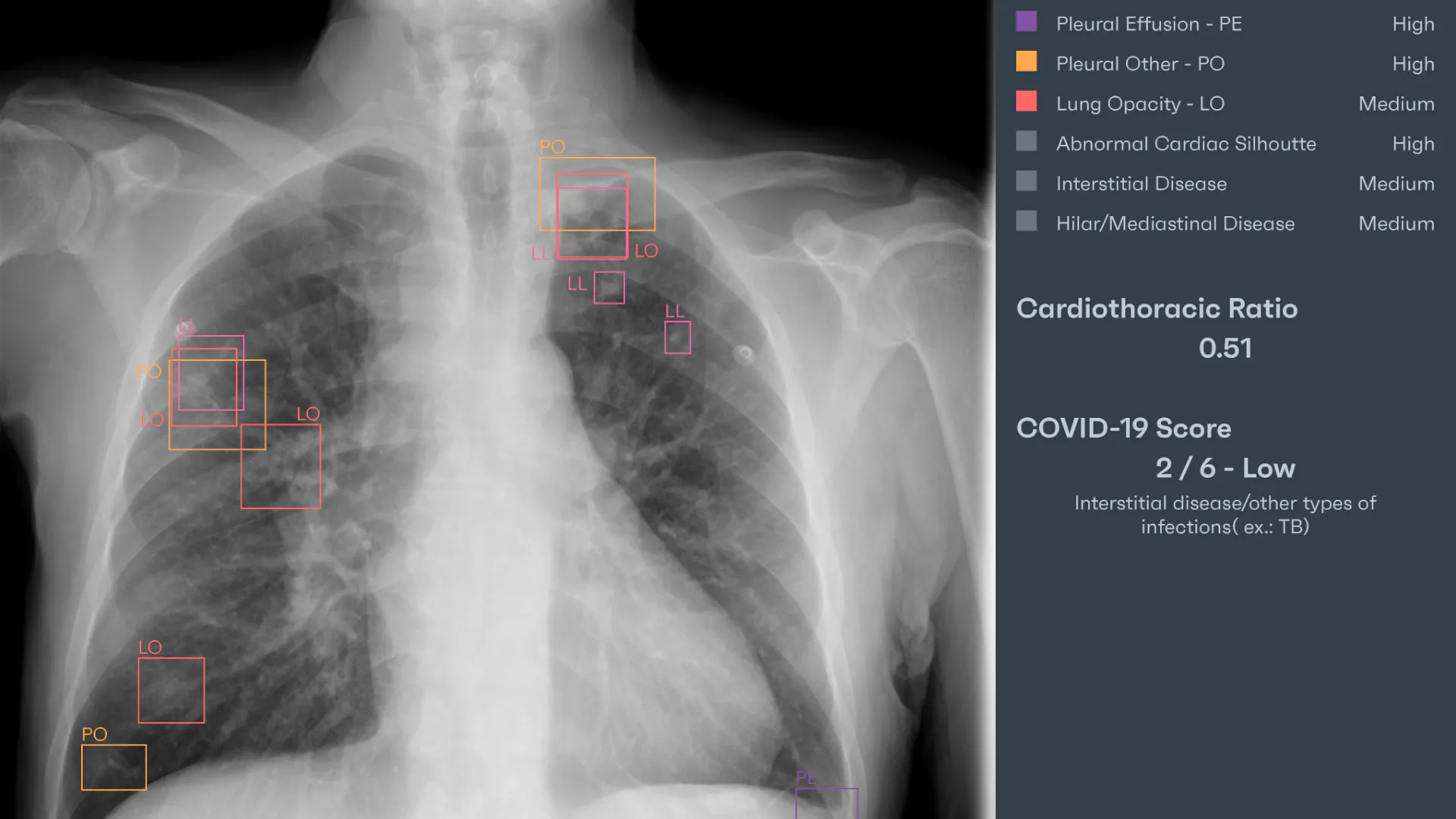
AI algorithms could automate routine tasks, such as identifying and tracking lesions, freeing up time for radiologists to focus on more complex cases. This could lead to a more efficient and streamlined radiology department, improving patient care and outcomes.
However, the implementation of AI radiology software must be approached with caution. There are concerns that overreliance on AI could lead to a reduction in human expertise and the potential for misdiagnosis. Furthermore, there are ethical considerations, including issues surrounding data privacy and bias in algorithm development. It is therefore crucial that any AI software is rigorously tested and validated before clinical use, and that radiologists and clinicians are adequately trained in its use.
Rayscape Artificial Intelligence: A potential solution to managing long-term sequelae in COVID-19 survivors
With Rayscape, we want to empower doctors with Artificial Intelligence tools for better decision-making.
We know that radiologists are constantly under pressure to make accurate diagnoses promptly, and our software was designed to help them be more, to be better. Rayscape's suite of products dedicated to the health of the lungs is seamlessly integrated into the doctor's and hospital's workflow, analyzing chest x-rays and lung CTs faster and more efficiently.

Rayscape CXR is automatically distinguishing between normal X-rays and those with anomalies, enhancing patient care and providing doctors with a quick way to triage and prioritize each patient. With over 147 pathologies that can be detected, our product has the amazing ability to bring an issue to light, eliminating doubt.
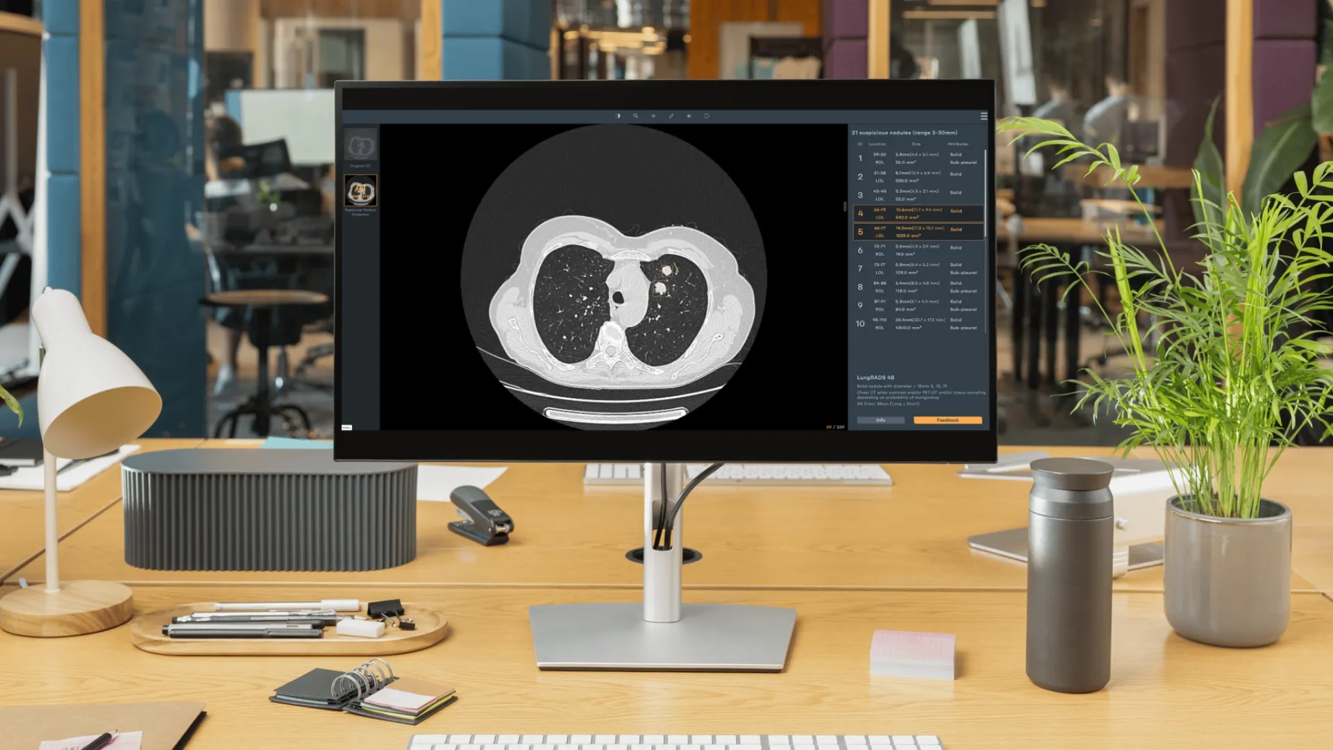
In addition, Rayscape Lung CT is an AI assistant that is trained in managing complex lung pathologies, from detecting and tracking lung nodules to evaluating COVID patients. Our product helps identify high-risk patients and notifies doctors by detecting possibly malignant nodules during regular check-ups, enabling seamless process optimization, from preventive monitoring to early detection, emergency care, and much more.
We understand that each hospital and clinic has unique needs, which is why Rayscape is flexible enough to plug into pre-existing infrastructures and workflows without disruptions. We offer a fully customizable platform, with both on-premise and cloud integrations, allowing hospitals and clinics to tailor Rayscape to their unique needs.
Reference:
Han, X., Chen, L., Fan, Y., Alwalid, O., Jia, X., Zheng, Y., Liu, J., Li, Y., Cao, Y., Gu, J., Liu, J., Zheng, C., Ye, Q., & Shi, H. (2023). Longitudinal Assessment of Chest CT Findings and Pulmonary Function in Patients after COVID-19. Radiology. https://doi.org/10.1148/radiol.222888


Discover how Rayscape works
integrate Rayscape AI into
your hospital's infrastructure
and improve both your
daily workflow and patients' experience.
