

Lung Cancer Screening with AI: Benefits and Challenges
Lung cancer is a significant global health issue, accounting for millions of deaths each year and costing billions of dollars in treatment. Early detection and treatment are essential to reducing mortality rates and costs but identifying tumors in their early stages can be a challenging task. While efforts have been made to detect lung cancer, the five-year survival rate for this cancer is still lower than that of other cancers, such as colorectal, breast, and prostate. However, if detected early, the five-year survival rate significantly improves.
Epidemiology of Lung Cancer
Worldwide, lung cancer is the second most commonly diagnosed cancer, and NSCLC (non-small cell lung cancer) is the most frequent type of lung cancer in the United States, accounting for 81% of all lung cancer diagnoses. In 2023, an estimated 238,340 adults (117,550 men and 120,790 women) in the United States will be diagnosed with lung cancer. Worldwide, an estimated 2,206,771 people were diagnosed with lung cancer in 2020, including both small cell lung cancer and NSCLC.
Since around 2006, the incidence rates in the United States have decreased by over 1% each year in women compared to 2.6% in men. This reduction in cases for both genders is attributed to fewer people smoking. The risk of lung cancer increases with age. An estimated 53% of all people diagnosed with the disease are age 70 or older, and an estimated 83% of cases are diagnosed in people age 65 or older. Men are most likely to be diagnosed with NSCLC between the ages of 80 and 84, while most cases in women are found between the ages of 75 and 79.
Lung cancer is the leading cause of cancer death for men and women worldwide. It is estimated that 127,070 deaths (67,160 men and 59,910 women) from this disease will occur in the United States in 2023. In 2020, an estimated 1,796,144 people died worldwide from the disease. Lung cancer accounts for approximately 20% of cancer deaths in the United States. However, death rates for the disease have declined by 58% since 1990 in men and 36% since 2002 in women through 2020. From 2014 to 2020, the death rates for men with lung cancer dropped by around 5% each year, and the death rates for women with lung cancer declined by 4% per year during the same period. Research suggests that these declines are due to fewer people starting smoking, more people quitting smoking, and advances in diagnosis and treatment.
Chest X-ray vs Lung CT
The most common screening method for detecting lung nodules, chest X-rays, is not always effective in identifying early-stage growths. More sensitive methods, such as low-dose computed tomography (CT), have been developed to provide more accurate depictions of anatomical structures, but they are not perfect. There is also a high degree of "observer error," where the lesion is missed during diagnosis, leading to delayed or missed treatments.
With the advent of artificial intelligence (AI) technology, there is an opportunity to provide a "second pair of eyes" to improve the accuracy of tumor detection and reduce the risk of observer error. By analyzing vast amounts of data, AI systems can identify patterns and changes that may indicate the presence of cancer, enabling earlier detection and treatment. This could have a significant impact on lung cancer treatment, reducing mortality rates and lowering the cost of care.

However, the question remains: is this the type of problem that AI is capable of tackling? While AI technology has advanced significantly in recent years, there are still technical challenges that must be overcome to develop reliable and accurate AI systems for lung cancer detection. The technology must be trained on large datasets of high-quality images to ensure accurate and consistent results. Additionally, AI systems must be integrated into clinical workflows and meet regulatory standards to ensure patient safety and privacy.
Identifying potential nodules, especially if they are in their early stages, is a very tough task. This is the main challenge of detecting cancer in X-rays and other radiology examinations.
Instead of being the best tools for the job, many of the first-line cancer screening instruments (like X-rays) are chosen because they are common and affordable. The task is extremely time-consuming, involves meticulous attention to detail over extended periods, and predominately includes imaging without malignancy. It must be carried out by highly qualified sub-specialty doctors.
Nevertheless, less sensitive techniques like low-dose CT scans have their restrictions and difficulties. Despite the aforementioned flaws, chest X-rays are still the primary line of screening for lung cancer due to their accessibility and affordability. One of the first things a doctor will order is a chest X-ray when a patient complains of chest pain, tiredness, or shortness of breath in an emergency department or clinic setting.
X-rays allow for the assessment of the heart, lungs, blood arteries, airways, and bones of the spine and chest in a single image. They are simple to obtain, inexpensive, and useful for ruling out a variety of illnesses that might be the source of the symptoms driving a patient to seek medical attention.
Sadly, there are considerable drawbacks to using X-rays to detect cancer:
- Limited sensitivity: X-rays have limited sensitivity in detecting small or early-stage lung cancers, as they can be easily obscured by other structures in the chest, such as ribs or the heart.
- Risk of false negatives: Radiologists may miss small nodules or misclassify them as benign, leading to a false-negative result.
- Radiation exposure: Although the amount of radiation used in a chest X-ray is relatively low, repeated exposure to radiation can increase the risk of cancer.
- Satisfaction of search: Radiologists may miss additional nodules or abnormalities if they become focused on a single finding, leading to a potentially missed diagnosis.
- Limited specificity: X-rays can produce false-positive results, leading to unnecessary follow-up testing and anxiety for the patient.
Given the limitations of X-ray, low-dose CT is now advised as an alternative for high-risk groups like ex-smokers. The main benefits are:

- Higher sensitivity: CT scans are more sensitive than X-rays, meaning they are better at detecting small nodules in the lungs that may indicate the presence of cancer. LDCT can detect nodules as small as 2-3 millimeters, while chest X-rays can only detect nodules that are several centimeters in size.
- More detailed images: CT scans provide more detailed images of the lungs and surrounding tissues than chest X-rays, which can help radiologists identify potentially cancerous nodules with greater accuracy.
- Lower radiation dose: While both LDCT and chest X-rays use ionizing radiation, LDCT uses a lower dose, reducing the risk of radiation-related health issues.
- Lower false-positive rates: LDCT has a lower false-positive rate than chest X-rays, which means it is less likely to detect nodules that are not cancerous, reducing the number of unnecessary follow-up tests and procedures.
- Greater survival benefit: Studies have shown that LDCT screening can reduce the risk of dying from lung cancer by up to 20% in high-risk populations compared to chest X-ray screening.
Several of the problems that prevent early detection in chest X-rays are also present in CT scans, despite being far more accurate for cancer screening (though at a lower rate). Radiologists misinterpret the results, miss significant findings, and give up too soon. This indicates that CT as a technique for early detection produces variable results: small, centrally placed nodules with ill-defined borders continue to be the most likely to be overlooked or to be mistakenly classified as benign.
Inter-grader variability is a problem in addition to instances where nodules are overlooked or scans are improperly interpreted. Even when radiologists concur that nodules are present, there are frequent disagreements on how to classify them.
Efficient Lung Cancer Detection
AI can revolutionize the way healthcare providers detect and diagnose lung cancer. The current challenge of being placed in front of 100 haystacks and told to determine which contains a needle is not a task well-suited for the human mind. This is where AI comes in; it excels in precisely this type of work.
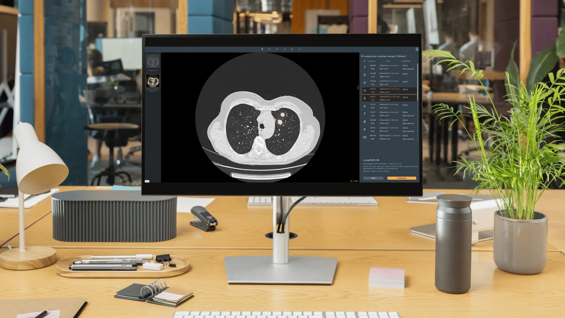
The need for early detection has fueled the development of scanning protocols and tools to detect even smaller lung tumors. While this is a good thing, it also requires more effort and attention from radiologists to read them. AI tools can help to shift some of the burden from busy and overloaded specialists to machines that don't become fatigued or distracted. One of the limitations of human vision is the ability to detect tiny cancerous tumors, leading to their being overlooked. AI tools can analyze multiple representations of data to highlight subtle patterns that might not yet be recognized as growth. This can help to detect small nodules that would otherwise be missed.
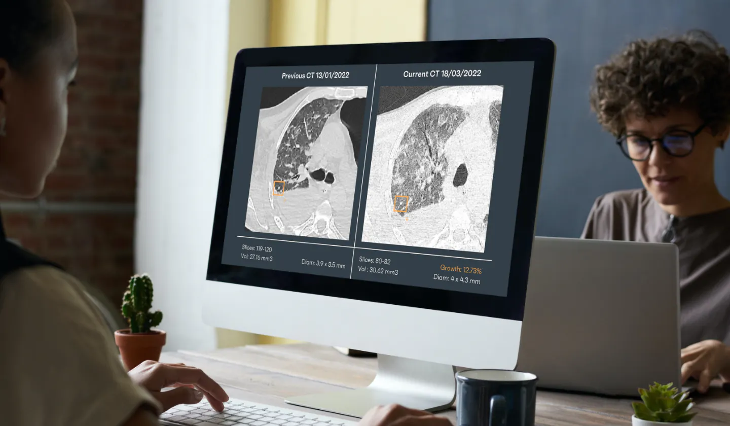
AI tools can also assist with more accurate classifications of suspicious lung nodules as malignant or benign. They can determine which features may be of significance and combine features from multiple models to provide estimates of confidence for informing clinical decision-making. This can lead to faster and more accurate diagnoses, ultimately saving lives.
Here are some examples of how AI can do this:
- AI can review large amounts of radiology scans to detect cancer nodules quickly and accurately. For instance, now multiple companies are working on detecting breast cancer with a high accuracy rate, sometimes higher than human radiologists' rate of 88.9%.
- AI tools can highlight subtle patterns that human vision may overlook, increasing the accuracy of cancer detection.
- AI can analyze multiple representations of data to better classify suspicious lung nodules as malignant or benign. A study by Stanford University researchers found that an AI system outperformed human radiologists in detecting lung nodules on CT scans, achieving an accuracy rate of 94% compared to humans' 75%.
- AI tools can provide estimates of confidence to inform clinical decision-making, increasing the accuracy and efficiency of a cancer diagnosis.
- AI can reduce the burden on busy and overloaded radiologists, allowing them to focus on more complex tasks that require human expertise. This can help improve patient care and reduce the risk of burnout among radiologists.
- AI can work tirelessly, without becoming fatigued or distracted, and can review large amounts of data quickly and accurately. This can help increase the efficiency of cancer detection and diagnosis, ultimately leading to better patient outcomes.
In addition, Artificial Intelligence can also assist with the tracking and monitoring of lung cancer progression over time. By analyzing data from multiple imaging studies, AI algorithms can detect changes in tumor size, shape, and density, and provide insights into the effectiveness of different treatment options. This can help clinicians make more informed decisions about patient care and improve patient outcomes.
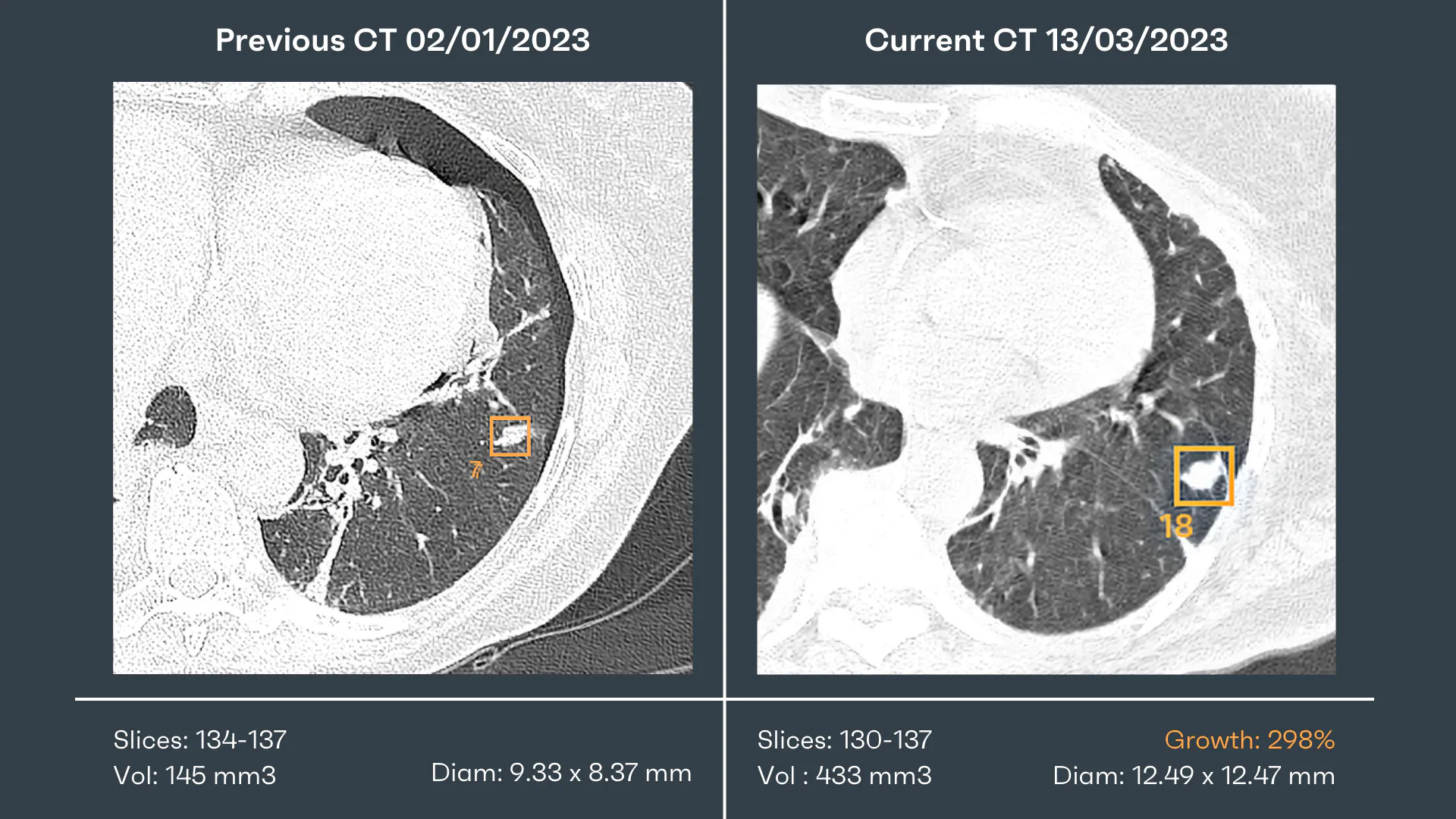
Lung Cancer Screening with Artificial Intelligence
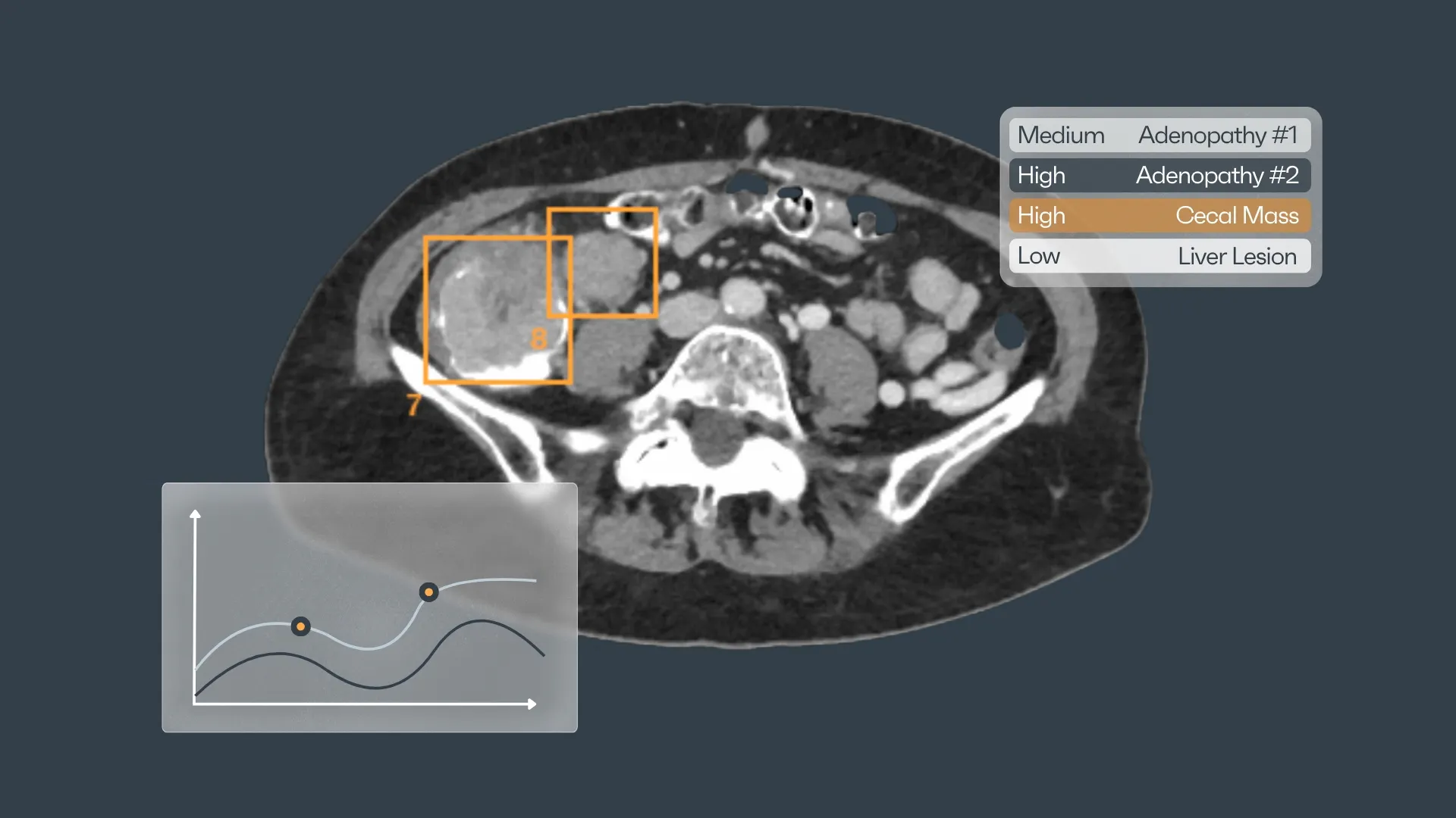
Moreover, Artificial Intelligence can also aid in the development of personalized treatment plans. By analyzing data from a patient's medical history, imaging studies, and other relevant sources, AI algorithms can identify biomarkers that may predict how a patient will respond to different treatments. This can help clinicians choose the most effective treatment for each patient, reducing the risk of complications and improving outcomes.
It can also play a role in improving the efficiency of clinical trials. By analyzing large amounts of data from diverse sources, including patient medical records, imaging studies, and genetic profiles, the software can identify patient cohorts that are most likely to benefit from a particular treatment. This can help streamline the clinical trial process and speed up the development of new cancer therapies.
Finally, with the help of Artificial Intelligence for lung cancer, we can also assist with the ongoing education and training of healthcare professionals. By providing access to large datasets and sophisticated analytical tools, we can help clinicians develop their skills and stay up-to-date with the latest advances in cancer diagnosis and treatment. This can help improve the quality of care delivered to patients and ultimately save lives.
So now, what should we do?
Here is what we think are the right steps to take:
- Start with a clear goal: The first step to implementing AI-powered cancer screening programs is to define clear goals and objectives. This means identifying the specific types of cancers to be targeted, the demographics to be served, and the desired outcomes.
- Invest in the right technology: Once the goals are defined, it's important to invest in the right technology to support AI-powered cancer screening. This includes hardware such as high-quality imaging equipment and software such as AI algorithms and machine learning tools.
- Collaborate with industry partners: Implementing AI-powered cancer screening programs requires a multi-disciplinary approach. Healthcare providers need to collaborate with industry partners such as technology companies, AI experts, and research institutions to develop effective screening programs.
- Train healthcare professionals: While AI can perform many tasks, it still requires human expertise to interpret the results and make informed decisions. As such, it's important to train healthcare professionals such as radiologists and oncologists in the use of AI-powered screening programs.
- Ensure data privacy and security: Implementing AI-powered cancer screening programs involves the collection and analysis of sensitive patient data. As such, it's important to ensure data privacy and security measures are in place to protect patient information.
- Monitor and evaluate outcomes: As with any healthcare program, it's important to monitor and evaluate the outcomes of AI-powered cancer screening programs. This includes assessing the accuracy of the AI algorithms, measuring patient outcomes, and identifying areas for improvement.
By following these steps, healthcare providers can implement AI-powered cancer screening programs that are effective, efficient, and provide better outcomes for patients.
References:
1. Cancer. [(accessed on 29 November 2021)]. Available online: https://www.who.int/news-room/fact-sheets/detail/cancer
2. Luo Y.H., Chiu C.H., Scott Kuo C.H., Chou T.Y., Yeh Y.C., Hsu H.S., Yen S.H., Wu Y.H., Yang J.C., Liao B.C., et al. Lung Cancer in the Republic of China. J. Thorac. Oncol. 2021;16:519–527. doi: 10.1016/j.jtho.2020.10.155. [PubMed] [CrossRef] [Google Scholar]
3. Cause of Death Statistics. [(accessed on 1 October 2021)]; Available online: https://www.mohw.gov.tw/lp-4650-2.html
4. Panunzio A., Sartori P. Lung Cancer and Radiological Imaging. Curr. Radiopharm. 2020;13:238–242. doi: 10.2174/1874471013666200523161849. [PMC free article] [PubMed] [CrossRef] [Google Scholar]
5. Migliore M., Palmucci S., Nardini M., Basile A. Imaging patterns of early-stage lung cancer for the thoracic surgeon. J. Thorac. Dis. 2020;12:3349–3356. doi: 10.21037/jtd.2020.02.61. [PMC free article] [PubMed] [CrossRef] [Google Scholar]
6. Travis W.D., Brambilla E., Nicholson A.G., Yatabe Y., Austin J.H.M., Beasley M.B., Chirieac L.R., Dacic S., Duhig E., Flieder D.B., et al. The 2015 World Health Organization Classification of Lung Tumors: Impact of Genetic, Clinical and Radiologic Advances Since the 2004 Classification. J. Thorac. Oncol. 2015;10:1243–1260. doi: 10.1097/JTO.0000000000000630. [PubMed] [CrossRef] [Google Scholar]
7. Nicholson A.G., Tsao M.S., Beasley M.B., Borczuk A.C., Brambilla E., Cooper W.A., Dacic S., Jain D., Kerr K.M., Lantuejoul S., et al. The 2021 WHO Classification of Lung Tumors: Impact of advances since 2015. J. Thorac. Oncol. 2021;17:362–387. doi: 10.1016/j.jtho.2021.11.003. [PubMed] [CrossRef] [Google Scholar]
8. Klang E. Deep learning and medical imaging. J. Thorac. Dis. 2018;10:1325–1328. doi: 10.21037/jtd.2018.02.76. [PMC free article] [PubMed] [CrossRef] [Google Scholar]


Discover how Rayscape works
integrate Rayscape AI into
your hospital's infrastructure
and improve both your
daily workflow and patients' experience.
