

Minimizing Radiology Errors: An Artificial Intelligence Guide
Reporting errors in radiology is an unfortunate reality. They're not intentional, and while some may have minimal impact on patients they carry significant consequences. The term "error" suggests a mistake, assuming there's always a clear-cut diagnosis and an infallible report. Yet, as radiologists, we know that imaging doesn't always yield a definitive outcome. Imaging outcomes aren't typically black and white, falling into the categories of "normal" or "abnormal." Our interpretations are based on our understanding of the patient's condition at the time of the exam. Often, what's deemed an "error" emerges later with additional information and a developing clinical picture.
That being said, the pursuit of minimizing reporting errors and enhancing accuracy remains crucial. Building artificial intelligence software specifically designed for radiology holds a wealth of knowledge and insights that can teach us invaluable lessons on reducing reporting errors. Through rigorous development and training, AI systems learn from vast amounts of data, enabling them to recognize patterns, identify abnormalities, and provide accurate assessments.
The process of constructing AI algorithms involves meticulous analysis of radiology images, comparing them against extensive databases of known cases. This iterative process allows the AI system to learn from both correct and incorrect interpretations, honing its abilities to make more accurate judgments over time.
Now, armed with a deeper understanding of the challenges inherent in radiology reporting and the transformative potential of artificial intelligence, we present to you our comprehensive guide. We will explore actionable strategies and practical steps that harness the power of AI to minimize reporting errors, enhance accuracy, and ultimately improve patient outcomes.

Step 1: Embrace Collaborative Approaches for Comprehensive Diagnoses
As a radiologist, it's crucial to embrace collaborative approaches. Working in conjunction with other healthcare professionals allows you to tap into their expertise and gain valuable insights that contribute to more accurate and holistic interpretations. Take the time to engage in discussions with referring physicians, surgeons, oncologists, and other specialists involved in the patient's care. By doing so, you'll gain a deeper understanding of the clinical history, symptoms, and treatment plans, which can greatly enhance your ability to evaluate imaging findings and make informed decisions for the patient's well-being.
Additionally, actively participating in multidisciplinary team meetings and boards can provide a unique opportunity. These platforms foster an environment where different perspectives can be shared and evaluated collectively. By leveraging the collective knowledge and experience of diverse healthcare professionals, you'll have the chance to refine your interpretations, improve diagnostic accuracy, and contribute to better patient outcomes.
Step 2: Reduce burnout and take breaks
Prioritize the most challenging cases right at the start of your shift, when your mind is at its peak freshness. Tackle those complex diagnostics head-on, leveraging your sharp focus and analytical prowess. By giving these cases your undivided attention early on, you set the stage for success and ensure the best possible outcomes for your patients.
Structured breaks are an absolute must. Take intentional pauses to recharge and reset. Step away from the screen, stretch those muscles, and give your mind a breather. Studies have shown that these strategic breaks improve cognitive function and combat mental fatigue.
Another valuable technique is periodically switching between modalities. By diversifying your workflow and alternating between different imaging techniques, you engage different cognitive processes. This not only prevents monotony but also enhances your overall performance. Embrace the versatility and adaptability that comes with navigating various modalities.
Now, let's address the elephant in the room: unnecessary interruptions and distractions. In a fast-paced radiology environment, it's all too easy to get pulled away from our core tasks. But we must take control of our environment. Streamline workflows, minimize interruptions, and create a focused space where distractions are kept to a minimum. Remember, our goal is precision, and that requires an undisturbed mindset.
Step 3: Master the Art of Effective Report Writing
Sometimes, radiologists may find themselves accurately interpreting imaging studies but falling short in effectively conveying their meaning through written reports. From the patient's perspective, the outcome can be the same whether a potential diagnosis is missed or if the relevant abnormalities are identified but poorly communicated in a report. Reports that are incoherent, rambling, and verbose, making it difficult to discern the most important findings, contribute to communication failures and can be as detrimental as missing relevant findings entirely.
While structured reporting may not be universally embraced, its benefits become evident, particularly for complex imaging studies, as it enhances thoroughness and accuracy. The recommendation is to prioritize simplicity and clarity in reports, eliminating typographical errors and focusing on including essential information while excluding irrelevant details. Concise and impactful reports facilitate comprehension for referrers and patients, ensuring that critical information is easily understood and enabling informed decision-making.

Understanding the context in which the imaging study is being requested is essential in providing valuable insights and actionable recommendations. Taking the time to discuss the appropriateness and justification of scans with referrers fosters a collaborative approach and ensures that the imaging study aligns with the clinical question at hand.
In addition, radiologists should actively seek missing snippets of history to augment their interpretation. Often, critical pieces of information are inadvertently omitted or overlooked during the imaging process. By proactively asking for appropriate missing details, you can paint a more comprehensive clinical picture and enhance the accuracy of their reports. This proactive approach demonstrates a commitment to thoroughness and a dedication to providing the most accurate and meaningful information to support patient care.
Lastly, radiologists must be cognizant of the potential impact of their language choices and report organization. Reports should strive for clarity and simplicity, avoiding unnecessary jargon or overly technical language that may hinder comprehension. Using a logical and structured format can facilitate efficient reading and comprehension, enabling referrers to quickly identify the key findings and their clinical implications.
Step 4: Harness the Power of Technology to Optimize Efficiency
In today's digital landscape, technology plays a significant role in optimizing radiology workflow and improving efficiency. As a radiologist, it's essential to leverage technological advancements to streamline your practice and enhance overall productivity. Integrating picture archiving and communication systems (PACS) and radiology information systems (RIS) can provide seamless access to patient data, images, and reports, enabling efficient communication and collaboration among healthcare providers.
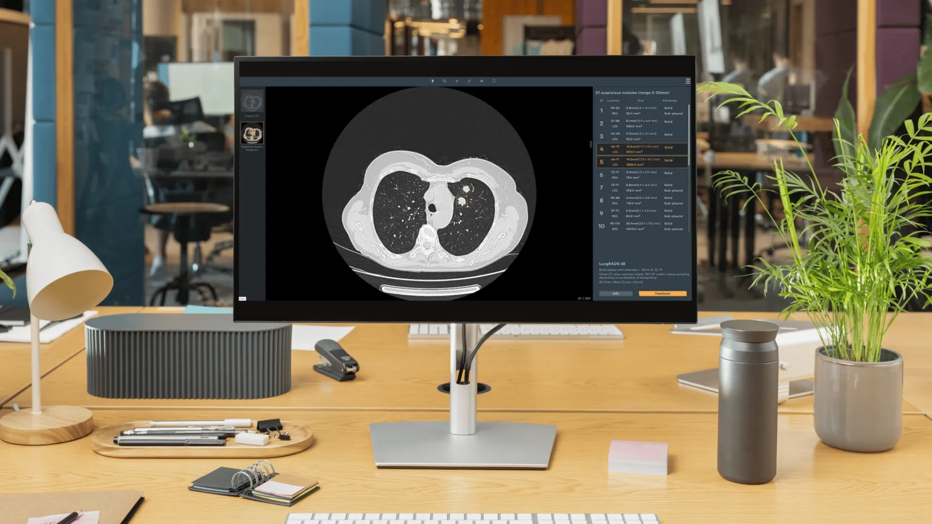
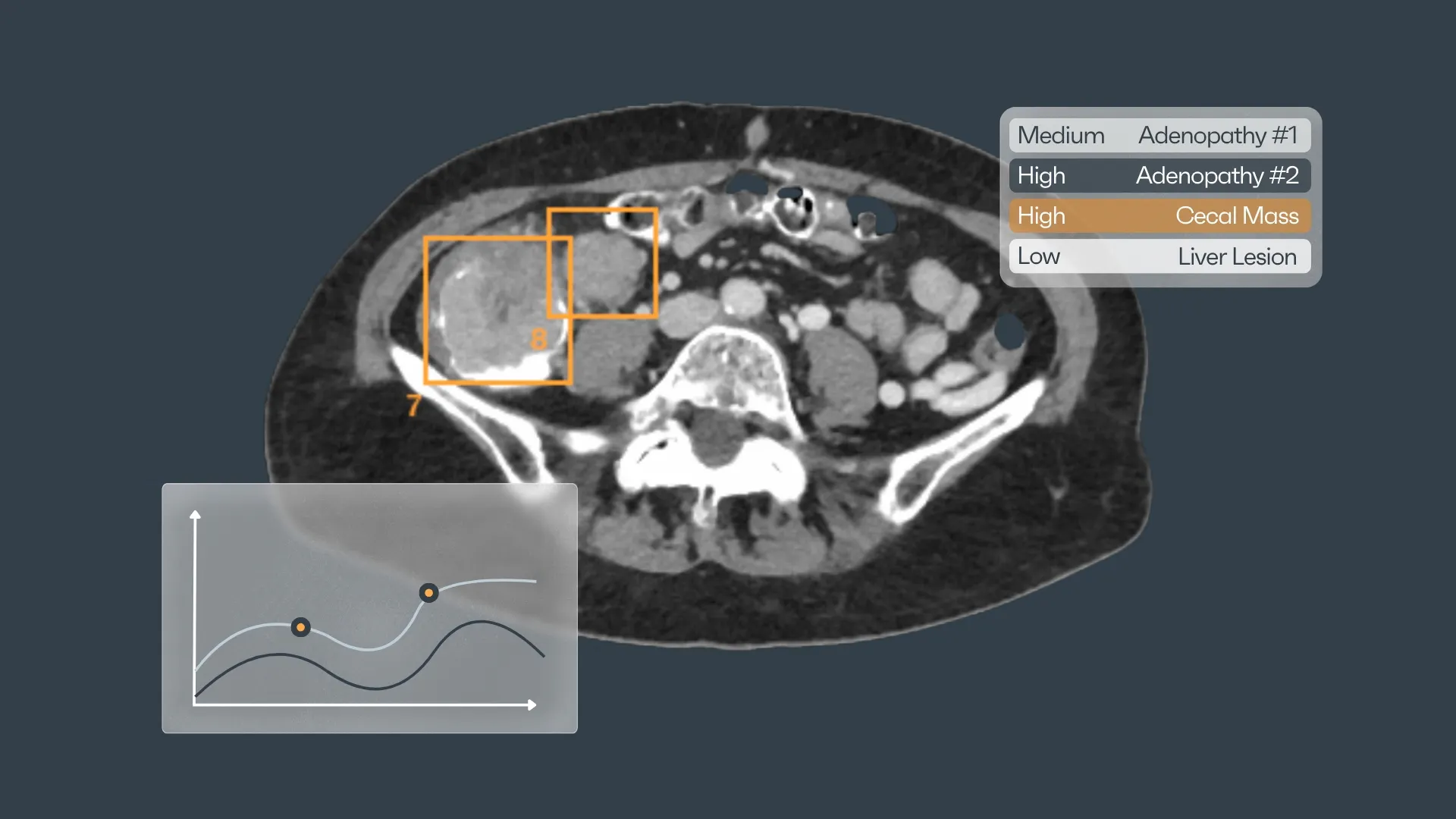
Furthermore, the integration of artificial intelligence (AI) and machine learning (ML) algorithms in radiology can revolutionize your interpretation process. These technologies have the potential to automate repetitive tasks, such as image segmentation and feature extraction, allowing you to focus on critical thinking and complex cases. AI and ML algorithms can analyze vast amounts of imaging data, detect subtle abnormalities, and assist in improving diagnostic accuracy while reducing interpretation time.
To stay ahead of the curve, explore emerging technologies like cloud-based solutions and telemedicine platforms. These innovations provide remote access to imaging data, facilitate remote consultations, and streamline communication with referring physicians. By embracing technology, you can transform your radiology practice, optimize efficiency, and ultimately deliver better patient care in the digital age.
Step 5: Unveiling Patient Insights for Enhanced Diagnostics
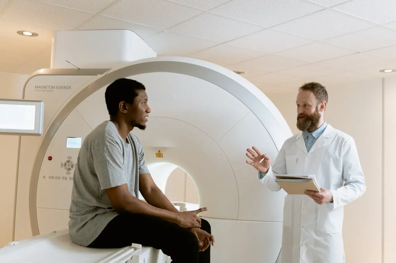
Finding time for clinical consultations with our referring colleagues and direct interaction with our patients can be a challenge. Numerous studies have shown that errors tend to creep in when offsite reporters, who have had no chance to interact with referrers or patients, are presented with limited clinical information. It's like trying to solve a puzzle with missing pieces. So, here's what we can do to tackle this issue head-on:
- Assess scan appropriateness and justification: We need to have those conversations about whether a scan is truly necessary and how it aligns with the clinical question at hand. It's about being strategic and not just ordering tests for the sake of it.
- Tailor studies to specific clinical questions: One size doesn't fit all in radiology. We should be tailoring our studies to match the specific information we need to answer the clinical question accurately. It's about precision and efficiency.
- Gather missing historical snippets: Don't be afraid to dig deeper and ask for those crucial pieces of information that might be missing. Time pressures may be real, but cutting corners won't do us any favors. Let's strive for comprehensive and complete reports.
- Engage in direct referrer discussions: Collaboration is key. Engage in direct discussions with referrers, whether it's one-on-one or within multidisciplinary team meetings. By understanding the clinical context and significance of the scan results, we can provide more accurate and meaningful reports.
Step 6: Overcoming Cognitive Biases in Radiology Practice
We all have cognitive biases. They're a product of our brains' attempts to simplify information processing. We can't completely eliminate these biases, but we must be aware of them and take corrective actions to minimize their impact on our reporting. Below are just a few of the many biases radiologists face, along with suggested measures to counter them.
- Anchoring bias: Sticking stubbornly to our initial impressions without considering new information. (Avoid premature guesses and strive to disprove your initial diagnosis instead of confirming it. Seeking a second opinion can sometimes be helpful.)
- Framing bias: Allowing the way a problem is presented to influence our interpretation. (Review the study blindly before reading the clinical information to avoid being swayed by framing.)
- Availability bias: Assigning a higher likelihood to a diagnosis simply because it comes to mind easily. (Rely on objective information to estimate the true base rate of the diagnosis rather than relying on quick impressions.)
- Satisfaction of search bias: Stopping the search for abnormalities once a likely diagnosis or initial abnormality is found. (Implement a systematic interpretation strategy, such as using a checklist or algorithm, to ensure a thorough review. Conduct a secondary search after detecting the initial abnormality and consider known combinations.)
- Premature closure bias: Accepting a diagnosis without complete verification. (Try to provide a differential diagnosis and never treat a working diagnosis as absolute without pathological confirmation.)

Remember, errors may occur despite our best efforts, but by adopting a proactive mindset, leveraging advancements in technology, and fostering collaboration, we can strive for accuracy and provide the best possible care for our patients.
In summary, radiologists should:
- Be aware of cognitive biases and take corrective actions to minimize their impact.
- Gather comprehensive patient information to support accurate diagnoses.
- Focus on improving report writing to effectively communicate findings.
- Manage mental and visual fatigue by prioritizing difficult cases and taking structured breaks.
- Embrace collaboration with colleagues and referrers to enhance diagnoses and treatment plans.
- Harness the power of technology, such as AI and digital tools, to optimize efficiency and accuracy.
- Stay connected with professional communities to stay informed and contribute to advancements in the field.
By implementing these strategies, radiologists can continuously improve their practice, reduce errors, and deliver better patient outcomes.
References:
- Berner ES, Graber ML. Overconfidence as a cause of diagnostic error in medicine. Am J Med 2008;121(5 suppl):S2–S23
- Wachter RM. Why diagnostic errors don’t get any respect: and what can be done about them. Health Aff (Millwood) 2010;29(9):1605–1610
- Abujudeh HH, Boland GW, Kaewlai R, et al. Abdominal and pelvic computed tomography (CT) interpretation: discrepancy rates among experienced radiologists. Eur Radiol 2010;20(8):1952–1957.
- Adrian Brady.Strategies to reduce reporting errors in radiology, European Congress of Radiology 2023
- Samei E, Krupinski E. Medical image perception. In: Samei E, Krupinski E, eds. The handbook of medical image perception and techniques. Cambridge, England: Cambridge University Press, 2010.
- Berlin L. Radiologic errors, past, present and future. Diagnosis 2014;1(1):79–84
- Revesz G, Kundel HL. Psychophysical studies of detection errors in chest radiology. Radiology 1977;123(3):559–562. Link, Google Scholar
- Siegle RL, Baram EM, Reuter SR, Clarke EA, Lancaster JL, McMahan CA. Rates of disagreement in imaging interpretation in a group of community hospitals. Acad Radiol 1998;5(3):148–154
- Donald JJ, Barnard SA. Common patterns in 558 diagnostic radiology errors. J Med Imaging Radiat Oncol 2012;56(2):173–178


Discover how Rayscape works
integrate Rayscape AI into
your hospital's infrastructure
and improve both your
daily workflow and patients' experience.
