

Does AI Use in Radiology Impact Lung Cancer Screening Programs?
Lung cancer is one of the deadliest cancers globally, responsible for 1.8 million deaths each year. Early detection remains a significant hurdle, often leading to diagnoses at advanced stages when treatment options are limited. Imagine a future where a quick scan not only detects lung cancer early but also pinpoints its exact location and progression in minutes. Many think about this being science fiction, but it isn't—it’s becoming reality with groundbreaking advancements in artificial intelligence made by both public and private organization pioneers.
Traditional lung cancer screening methods, such as chest X-rays and CT scans, have saved many lives but come with notable limitations, including high false-positive rates and difficulty detecting small or early-stage tumors. These challenges have driven the medical community to seek more reliable and efficient screening methods.
In this article, we will explore how AI is the next stage for lung cancer screening programs worldwide. We will highlight case studies from countries that have integrated new technologies into their healthcare systems, showcasing tangible benefits and challenges. Additionally, we will discuss the pioneering work of companies at the forefront of this technological revolution. By examining AI-enhanced radiology’s specific features and advantages, such as improved diagnostic accuracy, reduced screening times, and potential for personalized treatment plans, we aim to provide a comprehensive look at how AI redefines lung cancer detection.
Lung Cancer Screening: An Overview
Screening for cancer means catching it before any symptoms show up, which usually means it’s easier to treat. Early detection is a game-changer because finding cancer early on can significantly improve outcomes since it hasn’t had time to spread. Scientists are constantly researching who’s at risk and what factors contribute to developing cancer. This research helps guide who should get screened, what tests to use, and how often.

For lung cancer, there are three main screening methods:
1. LDCT (Low-Dose Computed Tomography): This uses low-dose radiation to take detailed pictures of your lungs. Studies, like the National Lung Screening Trial, have shown that LDCT can seriously cut down lung cancer deaths in heavy smokers by catching the disease early when it’s still treatable.
2. Chest X-rays: These are common, but they haven’t proven to be very effective in lowering lung cancer death rates.
3. Sputum Cytology: This involves checking the mucus you cough up for cancer cells. Like chest X-rays, it hasn’t been shown to reduce mortality effectively.
Screening for lung cancer has its upsides, but there are some risks too.
• False-Negative Results: You might get a negative result even if you have cancer, which could delay necessary treatment. Imagine getting a screening result that says you’re clear of cancer when, in fact, you have it. This is a false-negative—like a smoke alarm failing to go off during a fire, which can delay critical treatment.
• False-Positive Results: These can cause unnecessary stress and lead to more tests, like biopsies, which have their risks. Conversely, a false-positive result is when the test indicates cancer incorrectly, akin to a smoke alarm going off when it should not, causing unnecessary anxiety. These additional procedures carry also their own risks.
• Radiation Exposure: Both chest X-rays and LDCT expose you to radiation, which can increase cancer risk, especially in younger folks or those at lower risk.
Historical Context
The journey of AI in radiology started decades ago with computer-aided detection (CAD) systems helping radiologists spot abnormalities in medical images. Initially, these systems were pretty basic, often producing lots of false positives and limited by the computational power of the time. But as machine learning and neural networks advanced in the late 20th and early 21st centuries, AI began to not only detect but also diagnose various conditions with greater accuracy and efficiency.
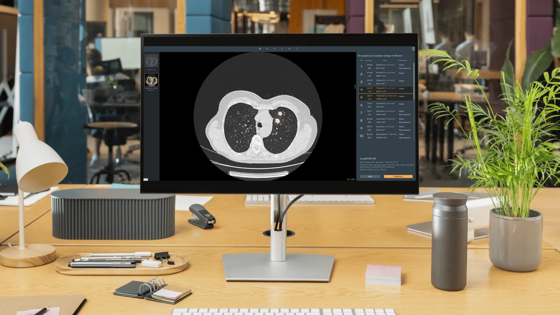
Current Technologies: Today, AI in radiology is incredibly advanced. Modern AI technologies can analyze complex medical images with remarkable precision, picking up subtle patterns and anomalies that the human eye might miss. For example, AI algorithms can now detect lung nodules, track their size and growth, and distinguish between benign and malignant lesions with high accuracy. They can also create enhanced images, like bone subtraction and suppression images, to help radiologists see abnormalities more clearly.
AI systems can even prioritize cases based on the severity of detected pathologies, ensuring urgent cases get prompt attention. This significantly cuts down diagnostic time and enhances early detection. By continuously learning from vast datasets, AI doesn’t just augment radiologists’ expertise—it brings a new level of precision and efficiency to medical imaging.
But that’s not all. AI technologies are also making strides in other areas. For instance, there’s tech that lowers radiation doses during scans, ensuring patient safety. There are also advanced algorithms that enhance image quality, making it easier to identify pathologies. On top of that, AI can detect patterns in clinical and biological information alongside medical imaging findings, giving a comprehensive view of a patient’s health.
Case Studies of Lung Cancer Screening Programs
Country Comparisons: Lung cancer screening programs vary widely across the globe. Up until 2021, Europe had just one established imaging screening program: mammography for breast cancer. But now, things are shifting. Low-dose computed tomography (LDCT) for lung cancer and magnetic resonance imaging (MRI) for prostate cancer are being rolled out, and discussions are ongoing about new imaging methods for breast cancer.
Back in the mid-1980s, the Europe Against Cancer program aimed to cut cancer mortality by 15% by 2000. By 2003, the EU was pushing for national screening programs for breast, cervical, and colorectal cancer. Fast forward to 2007, and many EU countries were starting or piloting these programs. By 2016, breast, cervical, and colorectal cancer screening was well-established across the EU.
In 2021, the European Commission launched Europe’s Beating Cancer Plan, with early detection as a key focus. This plan aims to broaden access to screening, bolstered by updated EU recommendations that expand the list of cancers to be screened from three to six, now including lung, prostate, and gastric cancers.
It’s a huge step forward, and with these new technologies and strategies, Europe is poised to make significant gains in the fight against cancer. Countries like Croatia, Poland, Italy, and Romania have committed to nationwide LDCT screening for high-risk individuals. Germany, the UK, and Sweden are conducting feasibility studies, while France, Belgium, the Netherlands, and Spain are cautiously running pilots and economic evaluations. Common challenges include governance, workforce capacity, and data management.
Success Stories: Poland has rolled out a centralized screening approach with regional centers and multidisciplinary teams, achieving impressive early detection rates. In the UK, the Targeted Lung Health Check program uses mobile CT scanners to reach underserved populations, boosting early diagnoses and participation.
Unique Approaches: Germany’s recent Lung Cancer Early Detection Ordinance emphasizes double reading by radiologists and AI integration, with mobile CT units enhancing accessibility. Italy focuses on centers of excellence to maintain high standards and consistency. The UK integrates screening with community services, particularly in deprived areas, demonstrating the effectiveness of localized healthcare interventions.
In the United States, LDCT screening is more widely adopted, driven by strong evidence from the National Lung Screening Trial and updated guidelines from the American Cancer Society (ACS). The ACS now recommends annual LDCT screening for adults aged 50-80 with a significant smoking history. This expanded eligibility is expected to save more lives by detecting lung cancer earlier.
In Japan, national screening programs utilize advanced AI to assist in reading scans, improving accuracy and reducing false positives. South Korea’s approach includes personalized screening protocols that leverage AI to assess individual risk factors, setting a new standard for precision in lung cancer detection.

The Role of Artificial Intelligence in Cancer Screening
We now understand that there is a perpetual need that technology needs to supply. Cancer screening, especially for breast, lung, and prostate heavily relies on imaging technology and, consequently, radiologists. This reliance means we need to consider the impact on radiologists’ workload and the rising burnout rates.
Radiologists are facing unmanageable workloads, leading to high burnout rates. For instance, a significant percentage of radiologists report feeling burned out, with the Medscape survey showing 54% experiencing burnout symptoms.
The study “AI-based pipeline for early screening of lung cancer: integrating radiology, clinical, and genomics data” published in The Lancet Regional Health - Southeast Asia explores a novel AI-driven approach to lung cancer screening.
Traditional methods like next-generation sequencing (NGS) have shown promise, but they face significant hurdles, particularly in resource-limited settings like India. NGS is costly and often hampered by issues like tissue adequacy. The study shines a light on a new frontier—leveraging AI to bridge these gaps.
Introducing the AI-Based Predictive System (AIPS): Imagine a system that can seamlessly integrate radiology, clinical data, and genomics to provide a comprehensive screening solution. That’s what the AI-based Predictive System (AIPS) aims to achieve. This AI-driven approach focuses on detecting and characterizing lung nodules from CT images and predicting the EGFR mutation status, which is pivotal for personalized treatment plans. The beauty of this system lies in its potential to democratize advanced screening methods, making them accessible and affordable even in low-resource environments.
Study Design and Data: The robustness of this study is rooted in its extensive dataset. Researchers analyzed data from 3,287 lung cancer patients across multiple cohorts, encompassing both Indian and White populations. By leveraging CT imaging and EGFR gene sequencing data, they trained and validated their AI models. This diverse dataset ensures that the findings are applicable to a broad range of populations, enhancing the study’s relevance and impact.
Performance of AIPS Models: The AIPS consists of two main modules: AIPS-N and AIPS-M.
• AIPS-N: This module is designed to detect and characterize lung nodules. It achieved an average precision of 70.19% in validation tests. This level of accuracy is significant because it demonstrates the system’s ability to reliably identify lung nodules, which is the first critical step in the screening process.
• AIPS-M: This module predicts the EGFR mutation status, a crucial factor in determining the most effective treatment plan for lung cancer patients. The AIPS-M module showed high accuracy with AUC values ranging from 0.79 to 0.91 across different cohorts. These results highlight the model’s robustness and its potential to be generalized across different populations.
Key Findings
The integration of AI with CT imaging is not just about automation; it’s about enhancing precision and efficiency in lung cancer screening. The study underscores several key points:
• Non-Invasive Prediction: The ability to non-invasively predict EGFR genotype through AI-driven analysis of CT images is a game-changer. It opens the door to more personalized treatment plans without the need for invasive procedures.
• Streamlined Process: By automating the detection and characterization of lung nodules, AI significantly reduces the need for manual annotation and complex feature engineering. This not only speeds up the screening process but also ensures consistency and accuracy.
The implications of this study are far-reaching:
• Improved Patient Survival Rates: It is clear by now that catching the disease in its early stages, treatment can be more effective, potentially saving lives.
• Optimized Resource Use: For healthcare systems, particularly in resource-limited settings, AI-driven models offer a cost-effective solution. They can help optimize the use of available resources, ensuring that advanced screening methods are accessible to a wider population.
• Global Impact: The study’s findings are not just applicable to India or the populations studied. They have the potential to influence lung cancer screening programs worldwide, making advanced diagnostic tools available to those who need them most.
Optimizing collaboration in lung imaging requires a different approach that involves both technological advancements and a cultural shift in the healthcare industry. In conclusion, to explore these diverse stages further and contribute to the evolving national programs, individuals can engage in ongoing education by:
- Using of Advanced AI Algorithms for Nodule Detection and Characterization: AI algorithms developed by organizations excel in detecting and characterizing lung nodules with high precision. These systems analyze CT scans to identify potential cancerous nodules, assessing their size, shape, volumetrics, densities, and expansion into adjacent tissues. This capability is crucial for early detection, as it can catch cancer in its nascent stages when treatment is most effective.
- Integrating Clinical and Genomic Data Analysis: Beyond just imaging, AI solutions integrate clinical and genomic data to provide a holistic view of a patient’s health. This integration allows the AI to predict genetic mutations such as EGFR, which are vital for personalized treatment plans. By combining these datasets, the system enhances the accuracy of its predictions and recommendations.
- Learning about Low-Dose CT Optimization for Radiation Minimization: Advanced AI technology optimizes the use of low-dose computed tomography (LDCT) scans. This is particularly important as it reduces radiation exposure for patients while maintaining high-quality imaging. However, the financial burden of implementing widespread screening programs remains a significant challenge for many countries. By cutting the dose and utilizing AI, costs can be reduced, potentially making these programs more feasible and accessible.
- Implementing Enhanced Image Quality through Novel Imaging Sequences: AI systems include advanced image processing tools that enhance the quality of CT scans. These tools can suppress noise, improve contrast, and highlight important features, making it easier for radiologists to identify abnormalities. Additionally, AI introduces novel imaging sequences that were not previously used, further improving diagnostic accuracy and patient outcomes.
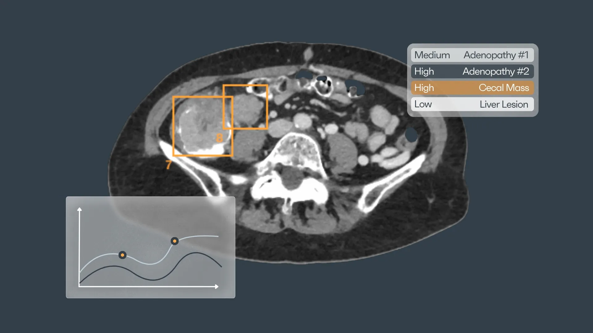
- Using Real-Time Analysis and Reporting Tools: A standout feature of AI is its ability to provide real-time analysis and reporting. Detecting cancer is just a small part of a radiologist’s job. Smart reporting and growth analysis give a better overview of what the patient has, translating into more personalized treatment plans. Immediate feedback allows for faster decision-making and quicker initiation of treatment plans, which is crucial in a time-sensitive disease like lung cancer.
- Automating the Workflows: AI streamlines the entire screening process by automating many of the routine tasks performed by radiologists. This not only increases efficiency but also reduces the burden on healthcare professionals, allowing them to focus on more complex cases and patient care.
- Understanding Predictive Analytics for Better Global Integration: AI systems include predictive analytics capabilities that can stratify patients based on their risk factors. With AI technology, we can now integrate not only local but also regional and global information into databases and statistics. This broad data integration allows new findings to emerge, potentially revolutionizing lung cancer screening and treatment approaches.
At Rayscape, we stand committed to pushing the boundaries of radiology management software, integrating the power of artificial intelligence to revolutionize patient care, enhance efficiency, and drive unprecedented advancements in the field of radiology. Join us on this transformative journey, where innovation meets excellence to shape the future of radiology.
Resources:
- https://www.thelancet.com/journals/lansea/article/PIIS2772-3682(24)00002-7/fulltext
- https://healthimaging.com/topics/healthcare-management/medical-practice-management/radiologists-rank-among-most-burned-out
- https://www.ilcn.org/lung-cancer-screening-in-poland-pilot-program-is-restarted-after-covid-19-lockdown/
- https://europeanlung.org/solace/2024/07/02/germany-introduces-first-steps-of-lung-cancer-screening-programme/
- https://www.lung.org/getmedia/186786b6-18c3-46a9-a7e7-810f3ce4deda/SOLC-2023-Print-Report.pdf#:~:text=URL%3A%20https%3A%2F%2Fwww.lung.org%2Fgetmedia%2F186786b6
- https://pressroom.cancer.org/releases?item=1274
- https://www.acr.org/Practice-Management-Quality-Informatics/ACR-Bulletin/Articles/July-2024/Burnout-Fueling-Workforce-Woes
- https://www.jtocrr.org/article/S2666-3643(22)00053-4/fulltext


Discover how Rayscape works
integrate Rayscape AI into
your hospital's infrastructure
and improve both your
daily workflow and patients' experience.
