

An in-depth look at AI Medical Image Processing in Radiology
Medical image analysis is a multifaceted discipline bridging computer science, mathematics, physics, and healthcare. It centers on the manipulation and interpretation of medical images to support diagnostics and therapeutic interventions. By examining these images, medical professionals gain noninvasive insights into human anatomy and physiology, enhancing their ability to detect, diagnose, and monitor diseases with greater precision.
At the heart of this field lies the application of computational techniques to extract essential diagnostic information from medical imagery. This involves several stages: image acquisition, preprocessing, segmentation, feature extraction, and classification. The ultimate objective is to elevate the quality and interpretability of medical images, thereby assisting clinicians in their decision-making processes.
The Medical Image Processing Pipeline
A standard workflow in medical image analysis consists of multiple critical steps, each contributing to the extraction of meaningful information for diagnosis or treatment:
1. Image Acquisition
The journey begins with the collection of medical images using various imaging modalities such as X-ray, magnetic resonance imaging (MRI), computed tomography (CT), ultrasound (US), and positron emission tomography (PET). The choice of modality depends on the clinical requirements, the area of the body being examined, and the specific diagnostic questions that need answering. High-quality image acquisition is crucial, as it sets the foundation for all subsequent processing and analysis steps.

2. Preprocessing
Once the images are acquired, they often require preprocessing to enhance their quality and prepare them for analysis. This stage involves:
- Noise Reduction: Eliminating artifacts and random variations in pixel intensity that can obscure important details.
- Contrast Enhancement: Adjusting the image to improve the visibility of features, making subtle differences more apparent.
- Standardization: Normalizing images to a common scale or format to ensure consistency across datasets, especially when combining images from different equipment or protocols.
Preprocessing aims to mitigate any imperfections introduced during image acquisition, such as motion blur or uneven illumination, ensuring that the images are in optimal condition for further analysis.
3. Image Segmentation
Segmentation is the process of partitioning an image into meaningful regions or segments to isolate areas of interest from the background. This step is pivotal for focusing analysis on specific anatomical structures or pathological regions, such as tumors, organs, or lesions. Techniques used in segmentation can be manual, semi-automated, or fully automated, leveraging algorithms like thresholding, edge detection, or advanced methods like convolutional neural networks (CNNs) in deep learning.
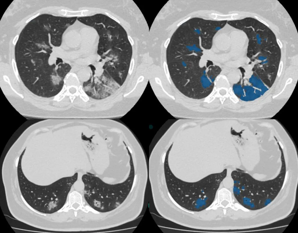
4. Feature Extraction
After segmentation, the next step is to quantify attributes or "features" of the identified regions. Features can include shape, texture, intensity, or spatial relationships and are essential for characterizing the regions of interest. In the context of deep learning, feature extraction is often performed implicitly by the layers of the neural network, which learns to identify relevant patterns and representations from the data automatically.
5. Prediction and Classification
Utilizing the extracted features, predictive models are employed to make clinical determinations. This could involve:
- Classification: Determining the category or class of the region, such as distinguishing between malignant and benign tumors.
- Regression: Estimating continuous variables, like the size of a lesion or the degree of disease progression.
- Detection: Identifying the presence or absence of specific conditions or abnormalities.
In deep learning frameworks, prediction and feature extraction are frequently integrated, with the network learning end-to-end mappings from input images to output predictions.
6. Post-Processing
Post-processing involves refining the predictions to enhance their accuracy and reliability. This may include:
- Filtering False Positives/Negatives: Applying thresholds or additional criteria to reduce errors.
- Smoothing Results: Adjusting prediction maps to eliminate spurious artifacts or noise.
- Validation: Comparing results against known standards or additional datasets to assess performance.
This stage ensures that the output is not only accurate but also clinically meaningful and ready for interpretation.
7. Visualization and Interpretation
The final stage focuses on presenting the results in a clear and accessible manner for clinicians. Effective visualization can involve:
- Overlaying Results: Superimposing segmentation masks or highlighting areas onto the original images to show the exact location of findings.
- 3D Reconstruction: Creating three-dimensional models from image slices to provide a comprehensive view of anatomical structures.
- Reporting Metrics: Providing confidence scores, measurements, or statistical summaries to aid in decision-making.
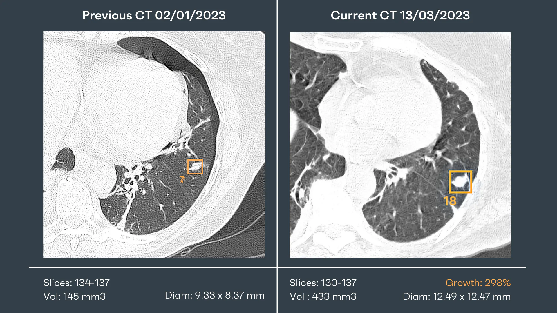
Visualization tools help bridge the gap between complex computational outputs and practical clinical insights, enabling physicians and radiologists to make informed decisions based on the analyzed data.
Preprocessing Techniques in Medical Imaging
Before applying AI algorithms or analytical techniques, medical imaging data undergo several preprocessing steps to ensure optimal performance:
- Image Normalization: Standardizing pixel intensities to reduce variability across images due to different scanning parameters, devices, or patient characteristics. Techniques include histogram equalization and z-score normalization.
- Noise Reduction: Employing methods like smoothing filters or anisotropic diffusion to eliminate artifacts introduced during image acquisition, thus enhancing image quality.
- Image Enhancement: Applying techniques to improve visual clarity and highlight features of interest, such as contrast stretching or edge enhancement.
- Image Resizing: Adjusting images to a consistent size and resolution to ensure uniformity across datasets, especially when sourced from various equipment or protocols.
- Image Augmentation: Expanding and diversifying the training dataset through rotations, scaling, flipping, and adding noise to improve AI model generalization. Advanced methods like generative adversarial networks (GANs) are also used to create synthetic images.
Each preprocessing step plays a vital role in enhancing the reliability and performance of medical image analysis, necessitating careful consideration based on the specific requirements of each task.
Practical Considerations and Future Directions
In implementing this pipeline, several practical aspects must be considered to ensure its effectiveness and integration into clinical workflows:
- Interoperability: Ensuring that systems and software can communicate seamlessly, often requiring adherence to standards like DICOM (Digital Imaging and Communications in Medicine) for image formats.
- Standardization: Establishing consistent protocols and methodologies across different institutions and equipment to facilitate data sharing and collaborative research.
- Artificial Intelligence Integration: Leveraging AI and machine learning, particularly deep learning, to automate and enhance various stages of the pipeline, from segmentation to prediction.
The Role of Artificial Intelligence
AI has become an indispensable component in modern medical image analysis. Its capabilities include:
- Automated Feature Learning: Deep learning models can learn complex representations directly from data without manual feature engineering.
- Improved Accuracy: AI algorithms have demonstrated high levels of accuracy in tasks like disease detection and classification, sometimes surpassing human experts.
- Efficiency Gains: Automation reduces the time required for analysis, enabling faster diagnosis and treatment planning.

Challenges and Ethical Considerations
While the integration of AI offers numerous benefits, it also presents challenges:
- Data Privacy: Ensuring patient confidentiality and compliance with regulations like HIPAA when handling medical images.
- Bias and Fairness: Avoid biases in AI models that can arise from unrepresentative training data, which may affect the generalizability of results across diverse populations.
- Explainability: Develop models whose decision-making processes are transparent and interpretable to gain the trust of clinicians.
Medical Imaging Modalities and the Impact of AI
Various imaging techniques offer distinct benefits and limitations. The choice among them depends on the clinical question, patient factors, and resource availability. Importantly, deep-learning AI techniques have proven effective in enhancing diagnostic accuracy across all five primary medical imaging modalities.
X-Ray Imaging
As the most accessible and cost-effective method, x-ray imaging is commonly used for initial evaluations, particularly of skeletal structures and the chest. Its effectiveness diminishes with soft tissue imaging. Deep-learning AI has improved diagnostic accuracy in X-rays for conditions like pneumonia and cancer.
Computed Tomography (CT) Scans
CT scans provide detailed images of internal organs and bones but involve higher radiation doses than standard X-rays. They are valuable for complex cases requiring high-resolution imaging but may not be suitable for patients who cannot tolerate contrast materials or need to limit radiation exposure. AI applications have enhanced CT diagnostics, especially in detecting small tumors.
Magnetic Resonance Imaging (MRI)
MRI offers superior soft tissue contrast, making it excellent for neurological, musculoskeletal, and cardiovascular imaging. Challenges include high costs, longer scan times, and incompatibility with certain implanted medical devices. Deep-learning AI has advanced MRI diagnostics, notably in detecting brain tumors and multiple sclerosis.
Ultrasound
Ultrasound is safe, radiation-free, and provides real-time dynamic imaging, ideal for obstetric and some cardiovascular applications. Image quality can be affected by the patient's body habitus or obstructions like air or bone. AI has been utilized to improve ultrasound diagnostics, particularly in identifying fetal anomalies and heart defects.
Positron Emission Tomography (PET) Scans
PET scans offer functional imaging primarily used in oncology for cancer diagnosis and monitoring but expose patients to radiation and are less effective for detailed anatomical assessments. Deep-learning AI has improved PET diagnostics, aiding in early-stage cancer detection.
Medical Image Analysis as a Specialized Field
Medical image analysis can be considered a specialized subset within image processing and computer vision, adapting and extending techniques from these areas to medical applications. While terminology may vary, the processes generally encompass:
- Image Processing: Operations where the input is an image and the output is a modified version of the image, such as denoising or sharpening.
- Image Analysis: Operations where the input is an image and the output is a labeled image highlighting specific regions, edges, or contours.
- Computer Vision: Operations where the input is an image used by algorithms to perform tasks like object detection, recognition, classification, and tracking, as well as answering questions related to the image's content.
Conclusion and Future Perspectives
Medical image analysis using AI has revolutionized healthcare by significantly improving the quality of diagnostics and treatment planning. From image acquisition to prediction and classification, each step of the medical image processing pipeline plays a pivotal role in providing actionable insights. As artificial intelligence continues to advance, its integration into medical imaging is poised to further increase accuracy and efficiency.
The field also faces challenges, such as ensuring data privacy and addressing biases in AI models. As these challenges are addressed, medical image analysis will remain a cornerstone in modern medicine, shaping the future of patient care.

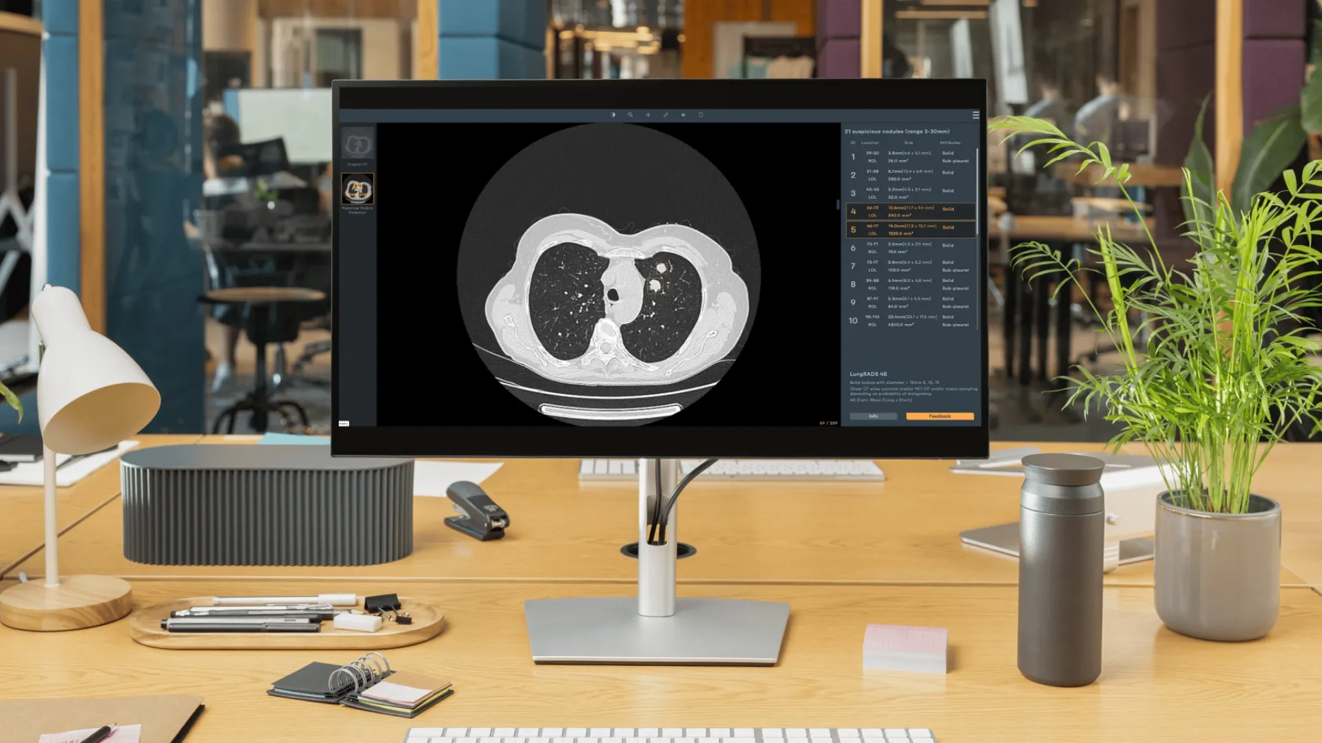
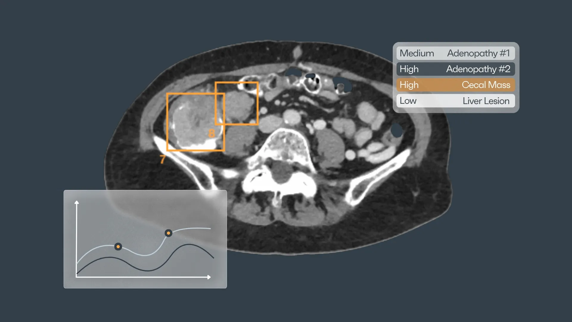

Discover how Rayscape works
integrate Rayscape AI into
your hospital's infrastructure
and improve both your
daily workflow and patients' experience.
