

Lung Cancer Screening with AI-Enhanced Pathway for CXR and CT
The journey to becoming a radiologist is a challenging yet rewarding path, demanding unwavering dedication, extensive education, and a passion for medical imaging. Radiology is a fascinating specialty that offers a diverse and dynamic workload, exposing professionals to a broad spectrum of pathologies and clinical scenarios. For those who enjoy solving complex puzzles, engaging in practical procedures, and contributing significantly to patient care, radiology presents an ideal career path.
In recent years, technological advancements have significantly transformed the field of radiology. The introduction of cutting-edge scanning equipment, advanced software tools, and innovative imaging techniques has been warmly embraced by the medical community. Modern medicine increasingly relies on imaging technologies to provide efficient, accurate, and non-invasive representations of diseases and illnesses. This reliance underscores the pivotal role radiologists play in diagnosing and managing a wide array of health conditions.
Introduction
One area where these technological advancements have made a profound impact is in the detection and management of lung cancer. Lung cancer remains one of the leading causes of cancer-related deaths worldwide. Early detection is crucial for improving patient outcomes, and radiologists are at the forefront of this battle. This article explores how artificial intelligence (AI) is revolutionizing lung cancer screening by integrating chest X-rays (CXR) and lung computed tomography (CT) scans. Let's understand how the superior lung cancer pathway that utilizes AI to enhance detection and monitoring, offers significant advantages over traditional methods.
The Evolution of Chest Radiography
The Historical Significance of Chest X-Rays
Chest X-rays have long been the cornerstone of respiratory assessment and are among the most frequently performed radiological examinations globally. Their widespread use is attributed to several factors:
- Cost-Effectiveness: Chest X-rays are relatively inexpensive compared to other imaging modalities, making them accessible for widespread use.
- Efficiency: They are quick to perform, providing immediate results that are critical in emergency and routine clinical settings.
- Simplicity: The procedure is straightforward, requiring minimal preparation, and can be performed on patients of all ages.
- Low Radiation Exposure: They involve minimal radiation dose, reducing the risk to patients and staff.
- Accessibility: Most medical facilities, including those in resource-limited settings, have the capability to perform chest X-rays.
For decades, the fundamental process of obtaining a chest X-ray remained largely unchanged. Radiologists relied on film-screen radiography systems, which, while effective, had limitations in image quality and diagnostic capabilities.
Transition to Digital Radiography
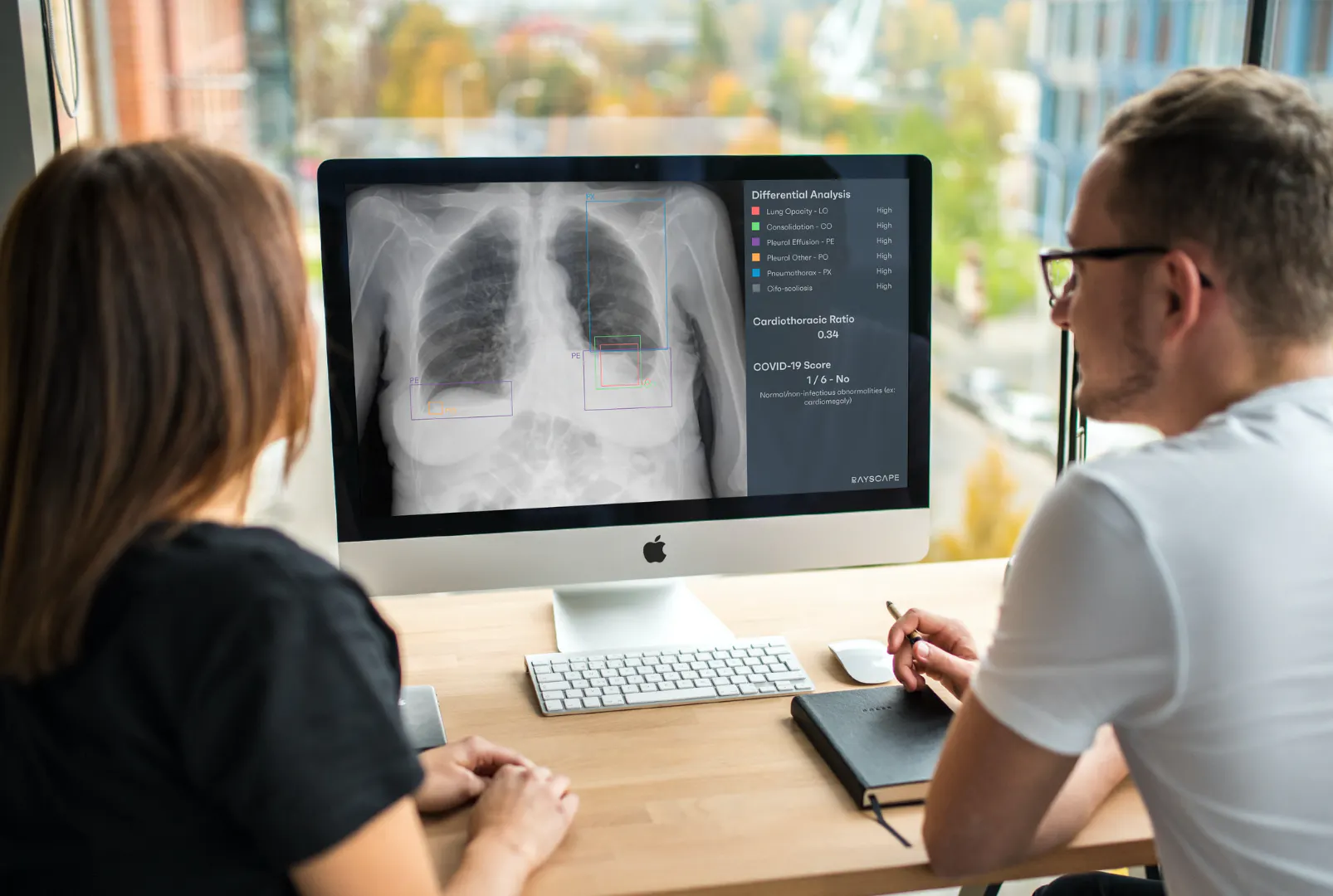
The advent of digital radiology has transformed the landscape of chest imaging. Exponential advances in processing power and digital technology have facilitated:
- Enhanced Image Quality: Digital detectors provide superior image resolution and contrast, enabling better visualization of anatomical structures.
- Image Manipulation: Radiologists can adjust image parameters such as brightness, contrast, and zoom, aiding in the detection of subtle abnormalities.
- Efficient Workflow: Digital images are readily available for review, eliminating the need for film processing and reducing turnaround times.
- Storage and Sharing: Digital images can be stored in Picture Archiving and Communication Systems (PACS), allowing for easy retrieval and sharing among healthcare professionals.
Advancements in Imaging Techniques
Recent advancements have further enhanced chest radiography:
- Dual-Energy Imaging: Separates images into different energy spectrums, improving tissue contrast and detection of lesions.
- Tomosynthesis: Provides quasi-three-dimensional imaging by acquiring multiple projections, reducing the overlap of anatomical structures.
- Computer-Aided Detection (CAD): Assists radiologists by highlighting areas of interest, increasing sensitivity in detecting abnormalities.

Challenges in Lung Cancer Detection
The Complexity of Interpreting Images
Despite technological advancements, interpreting chest images remains a complex task. Overlapping anatomical structures, such as ribs, clavicles, and pulmonary vasculature, can mask subtle findings, particularly in the mediastinum and lung fields. Variations in patient anatomy and positioning can further complicate interpretation.
Radiologists must be meticulous in their examination, following a systematic approach to ensure no area is overlooked. Common challenges include:
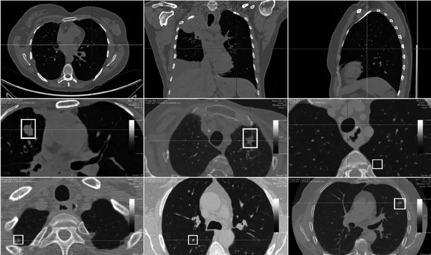
- Subtle Lesions: Small nodules or early-stage tumors may be difficult to detect due to their size and the complexity of the surrounding anatomy.
- Variability in Presentation: Lung cancer can present in various forms, including masses, infiltrates, or as part of complex patterns like interstitial lung disease.
- Perceptual Errors: Human factors such as fatigue, high workload, and cognitive biases can lead to missed diagnoses.
The Importance of Early Detection
Lung cancer is often diagnosed at an advanced stage due to the absence of symptoms in early disease. Early detection is critical for improving survival rates, as treatment options are more effective when the disease is localized. Small pulmonary nodules detected on imaging may represent early-stage cancer, necessitating careful evaluation and follow-up.
Traditional Methods of Nodule Detection
Traditionally, the detection and classification of lung nodules have relied heavily on the radiologist's expertise and experience. The process involves:
- Visual Assessment: Identifying nodules based on size, shape, density, and location.
- Comparison with Previous Studies: Evaluating changes over time to assess growth patterns.
- Risk Stratification: Considering patient factors such as age, smoking history, and exposure to carcinogens.
But, even with meticulous analysis, there is a risk of missing subtle or ambiguous findings, which can delay diagnosis and treatment.
The Superior Lung Cancer Pathway: Integrating AI with CXR and CT Scans
The Role of Artificial Intelligence in Radiology
Artificial intelligence has emerged as a transformative tool in radiology, particularly in the field of thoracic imaging. AI systems, powered by machine learning algorithms and deep neural networks, can analyze vast amounts of imaging data, identifying patterns and features that may be imperceptible to the human eye.

AI excels in several areas critical to lung cancer detection:
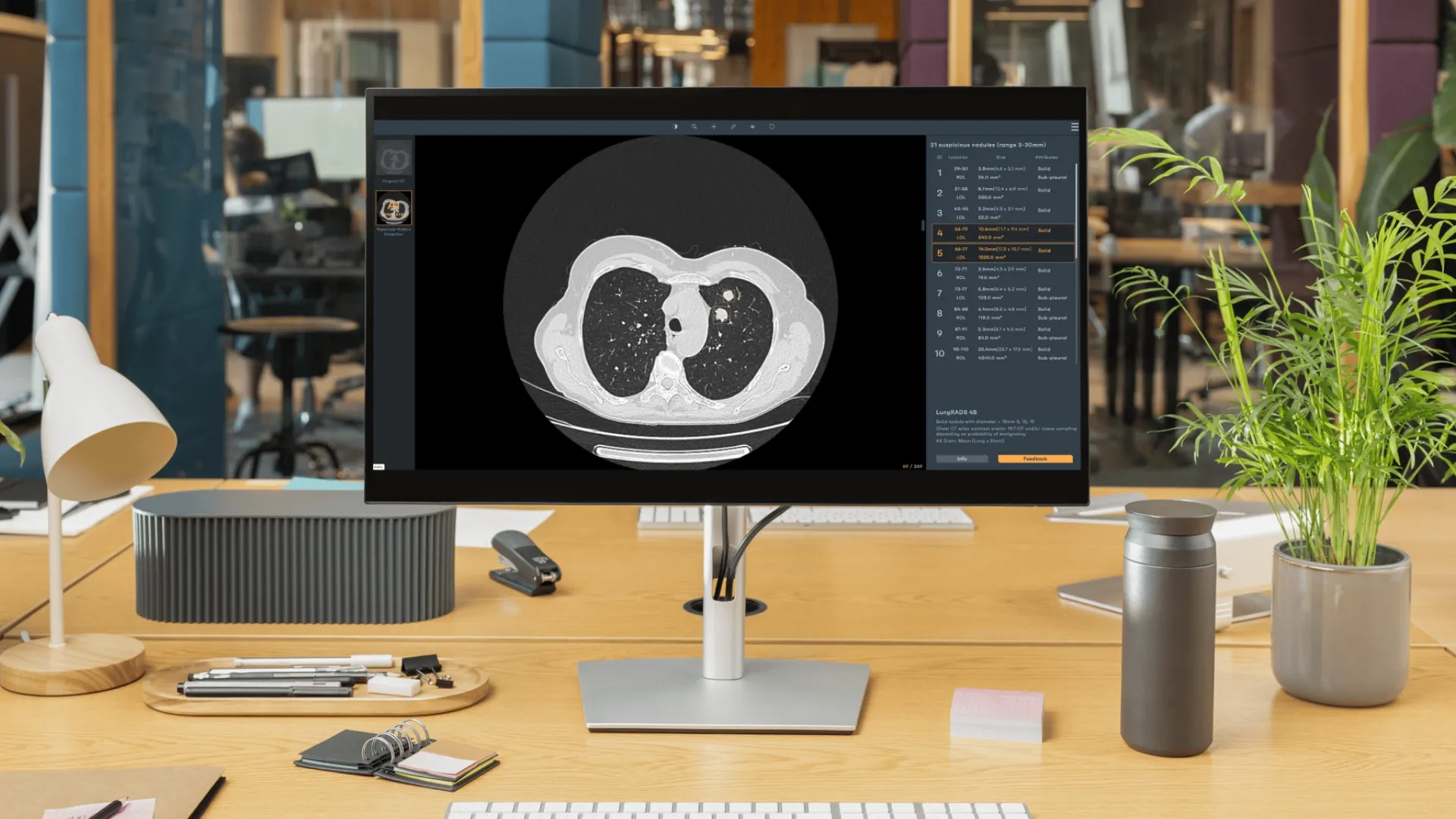
- Automated Nodule Detection: AI algorithms can automatically identify lung nodules on both chest X-rays and CT scans with high sensitivity and specificity.
- Nodule Characterization: AI assesses nodules based on size, shape, margin characteristics, internal structure, and attenuation patterns.
- Risk Assessment: Algorithms incorporate clinical data to provide a probability of malignancy, aiding in decision-making.
Combining CXR and CT Scans for Comprehensive Evaluation
Integrating AI with both chest X-rays and lung CT scans offers a synergistic approach:
- Initial Screening with CXR: Chest X-rays serve as a primary screening tool due to their accessibility. AI enhances interpretation by detecting subtle abnormalities that may be missed during routine analysis.
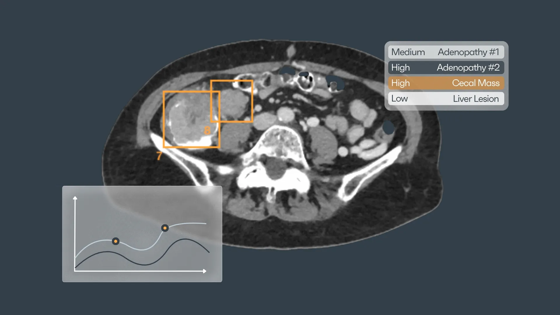
- Detailed Assessment with CT Scans: When abnormalities are detected on CXR, CT scans provide detailed cross-sectional imaging. AI algorithms further analyze CT data to evaluate nodule characteristics, assess the extent of disease, and identify lymphadenopathy or metastatic lesions.
This integrated pathway ensures that patients receive a thorough evaluation, with AI assisting at each stage to enhance accuracy and efficiency.
Benefits of the AI-Integrated Lung Cancer Pathway
Improved Diagnostic Accuracy
The AI-integrated pathway offers several advantages:
- Standardization: AI provides consistent analysis, reducing inter-observer variability among radiologists.
- Enhanced Sensitivity: Increases the detection rate of small and subtle nodules.
- Reduced False Negatives: Minimizes the risk of missed diagnoses, leading to earlier intervention.
Efficiency in Clinical Workflow
AI integration streamlines radiology services:
- Rapid Analysis: AI processes imaging studies quickly, providing preliminary findings that expedite diagnosis.
- Workload Reduction: By handling routine assessments, AI allows radiologists to focus on complex cases and consultative roles.
- Decision Support: AI offers evidence-based recommendations, supporting radiologists in clinical decision-making.
Personalized Patient Care
The AI pathway facilitates individualized treatment plans:
- Risk Stratification: Incorporates patient demographics, clinical history, and imaging findings to assess cancer risk.
- Monitoring and Follow-Up: AI tracks changes in nodules over time, aiding in surveillance strategies.
- Patient Engagement: Clear and precise imaging results enhance patient understanding, improving compliance with follow-up recommendations.
While AI offers significant benefits in lung cancer screening, several challenges remain that need to be addressed to fully realize its potential. One of the primary concerns is data quality and diversity. AI models require high-quality, diverse datasets for training to ensure accuracy across different populations. Without representative data from various demographics, the AI algorithms may not perform effectively for all patient groups, potentially leading to disparities in diagnosis and treatment.
Another significant challenge is integration and interoperability with existing hospital information systems. Seamless integration of AI solutions into current infrastructures like Picture Archiving and Communication Systems (PACS) and Radiology Information Systems (RIS) is essential. Without compatibility, the implementation of AI can disrupt workflows and reduce efficiency, hindering its acceptance among healthcare professionals.

Regulatory and ethical considerations also pose challenges. Compliance with healthcare regulations is necessary to ensure patient safety and data integrity. Addressing ethical concerns around AI usage, such as patient consent and algorithmic transparency, is crucial. Ensuring data security and confidentiality in line with regulations like HIPAA and GDPR protects patient privacy and fosters trust in AI applications.
Overcoming Challenges and Ensuring Success
To surmount these obstacles, a strategic approach is required. Ensuring data quality involves establishing rigorous protocols for data collection, labeling, and annotation. This improves the AI training process by providing accurate and consistent information. Incorporating data from a wide range of demographics ensures that the AI system is applicable across diverse populations, enhancing its reliability and effectiveness.
Integration with clinical systems demands collaboration with technology vendors to ensure AI solutions are compatible with existing hospital infrastructures. Providing comprehensive training for radiologists and staff on AI functionalities and workflows is essential. Educated users are more likely to embrace AI tools, utilizing them effectively to enhance patient care.
Addressing regulatory compliance and ethical use is paramount. Adhering to guidelines from regulatory bodies like the FDA and EMA ensures that AI applications meet required safety and efficacy standards. Maintaining patient privacy by securing data in compliance with HIPAA and GDPR regulations is critical. Transparency about AI use in diagnostics, communicated openly with both clinicians and patients, promotes trust and ethical responsibility.
Future Perspectives
Looking ahead, advancements in AI technology promise to further revolutionize lung cancer screening and radiology. The development of continuous learning models means AI systems can evolve by incorporating new data, continually improving their diagnostic capabilities over time. This adaptability enhances the precision and accuracy of lung cancer detection.
The integration of AI with Electronic Health Records (EHRs) represents another significant advancement. By combining imaging data with comprehensive clinical information, AI can provide a more holistic analysis of a patient's health, leading to better-informed decision-making and personalized treatment plans. Predictive analytics is an emerging field where AI models can forecast disease progression and treatment responses, offering proactive insights into patient care.
Expansion into other imaging modalities is also on the horizon. In molecular imaging, AI can assist in interpreting PET and SPECT scans, which are vital for assessing metabolic activity and tumor behavior. Advanced AI algorithms for lung MRI are particularly beneficial for patients where radiation exposure is a concern, providing detailed imaging without the associated risks of ionizing radiation.
Interdisciplinary collaboration is essential to drive these advancements. Partnerships between radiologists and engineers facilitate the development of AI tools that are tailored to clinical needs, ensuring practicality and effectiveness in real-world settings. Global health initiatives can leverage AI to improve lung cancer screening in resource-limited areas, reducing healthcare disparities by providing advanced diagnostic capabilities where they are needed most.

How to test out AI solutions today?
Rayscape is a European-based company specializing in AI-driven solutions to enhance radiologists' ability to analyze chest X-rays and lung CT scans. Our solutions, powered by 42 million medical images, have been deployed in national TB screening programs, aided doctors during the COVID-19 pandemic, and are used in ICU and Emergency Room imaging.
Expanding into oncology, Rayscape’s AI covers the full lung cancer pathway, ensuring early detection, accurate monitoring, and treatment support.
Recognizing the challenges of AI generalization early, we've focused on ensuring our algorithms perform well across all populations, through partnerships with medical institutions worldwide.
Try Rayscape today at: https://demo.rayscape.ai/home
Resources:
1. National Cancer Institute
Lung Cancer Screening (PDQ®)–Health Professional Version.
https://www.cancer.gov/types/lung/hp/lung-screening-pdq
2. National Lung Screening Trial Research Team
"Reduced Lung-Cancer Mortality with Low-Dose Computed Tomographic Screening."
New England Journal of Medicine, vol. 365, no. 5, 2011, pp. 395–409.
DOI: 10.1056/NEJMoa1102873
https://www.nejm.org/doi/full/10.1056/NEJMoa1102873
3. American Cancer Society
Key Statistics for Lung Cancer.
https://www.cancer.org/cancer/lung-cancer/about/key-statistics.html


Discover how Rayscape works
integrate Rayscape AI into
your hospital's infrastructure
and improve both your
daily workflow and patients' experience.
