

Why oncology CT imaging needs a new generation of AI
We've been watching AI transform radiology for years now, and it's been genuinely exciting to see how far we've come. The early tools, the ones that could spot lung nodules on chest CTs or identify brain hemorrhages with impressive precision, are all remarkable achievements. Each one represented a real breakthrough in what technology could do for patient care.
But as we've gotten deeper into this space, we've started thinking more about where we need to go next. And we keep coming back to this: we've gotten really good at analyzing individual organs, but patients aren't collections of organs. They're integrated systems. And nowhere is this more obvious than in oncology imaging.
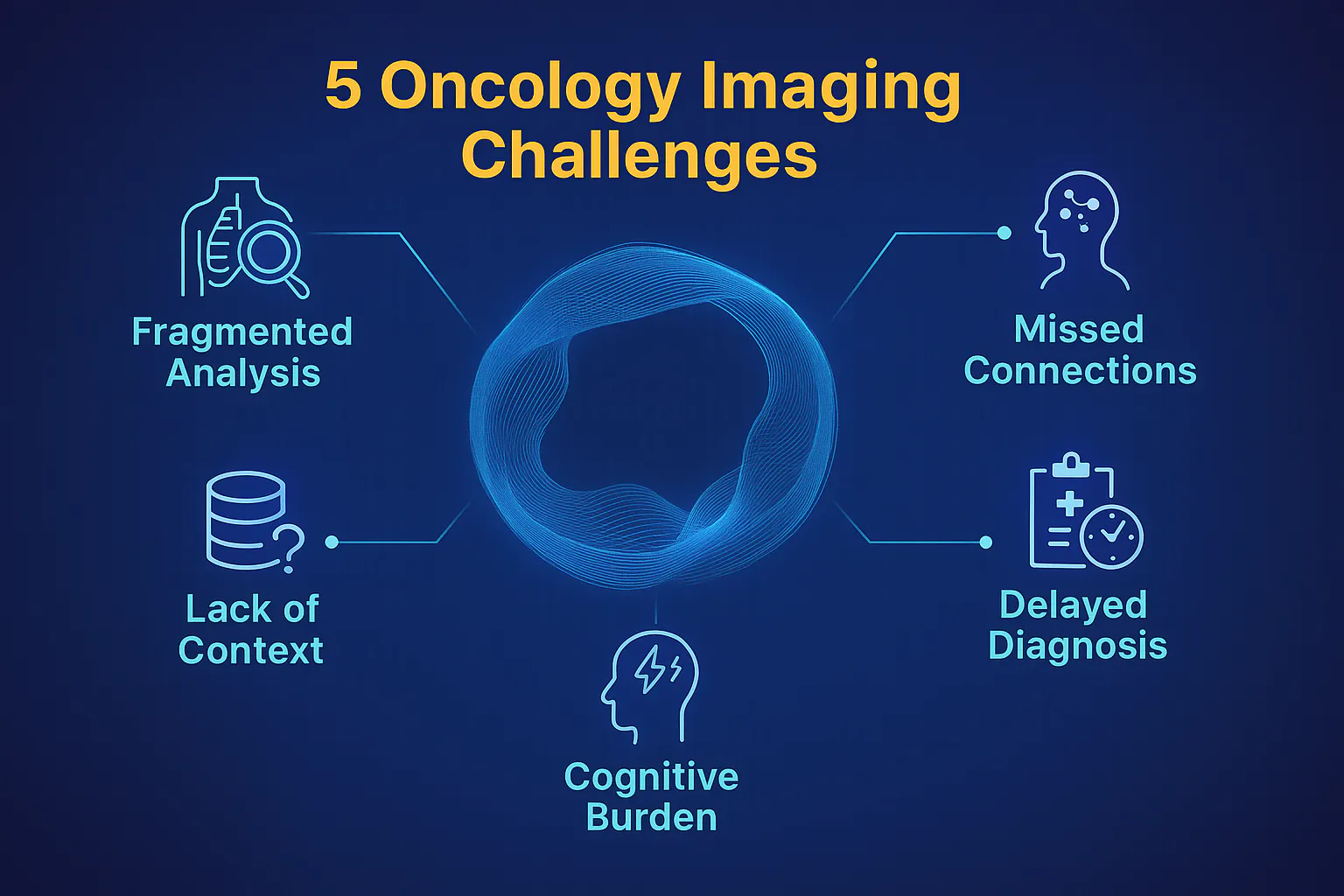
Here's what strikes us about where we are right now. Current tools are remarkable in their focus, but their fragmentation creates blind spots. Traditional “AI” platforms, if we can refer as that, focus on isolated organs, which results in fragmented diagnostics and missed correlations. There are incredible solutions for quantification of COVID lesions, detection of emphysema, alerting about vertebral fractures, etc; the precision is genuinely impressive, but something still feels like it’s missing. AI should be driven by multi‑organ imaging; it should be able to analyze several anatomical regions simultaneously and thereby “bridge diagnostic gaps”, as a holistic grasp of disease processes that transcend organ boundaries.
Fragmented intelligence: the limitations of point solutions
But in the day-to-day work as radiologists, doctors find themselves thinking about the next step. Because when we’re reading an oncology case, we are not just checking individual organs. We are looking for connections, patterns that span across anatomical boundaries. That's how disease actually behaves.
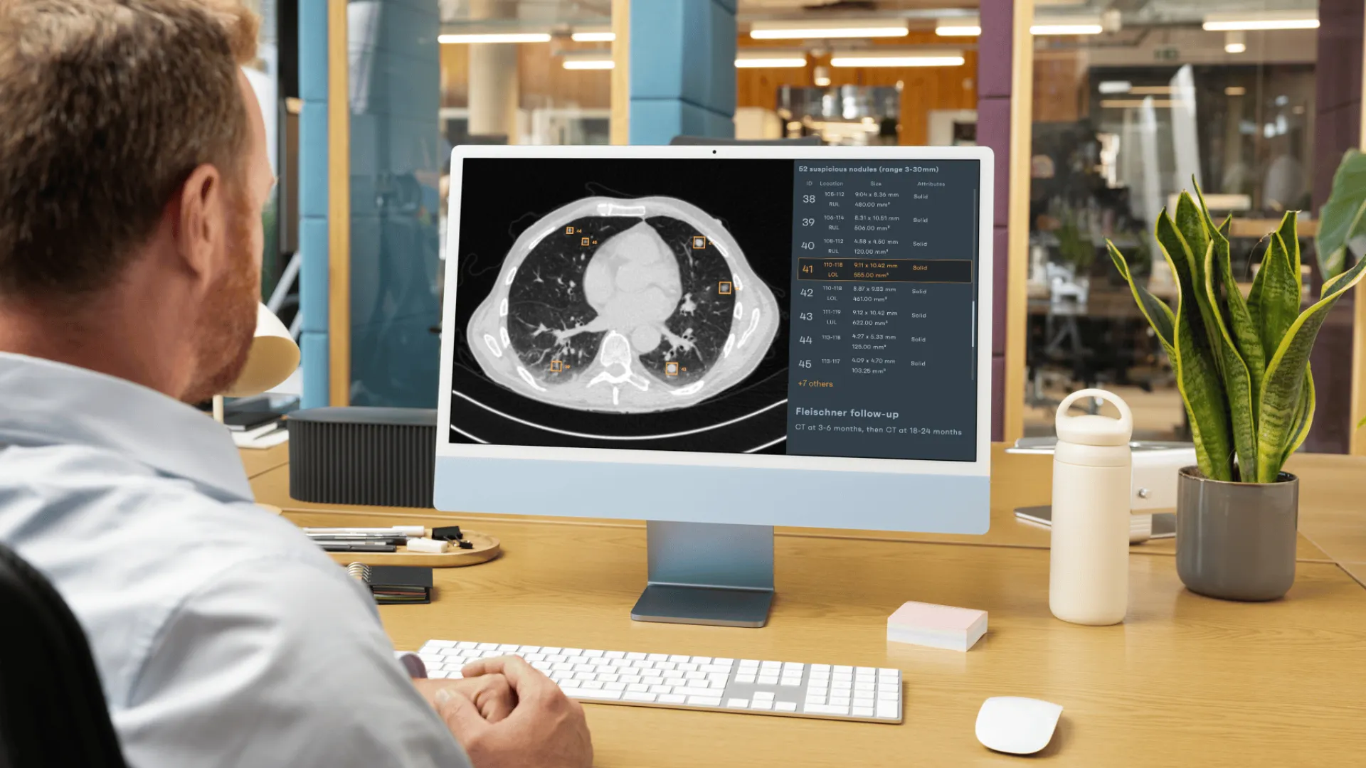
We'll be looking at a chest CT and notice a subtle nodule, and our brain immediately starts asking questions: Is there anything in the abdomen that could be related? What did the pelvis show? These connections matter. A pulmonary nodule might be related to hepatic lesions. Lymph node enlargement in one region might signal something happening elsewhere.
The challenge isn't that current tools can't detect these findings; they absolutely can. The opportunity is in helping radiologists connect the dots more seamlessly. Now, AI solutions address a narrow clinical need, yet together they present an overwhelming mosaic of dashboards and outputs that clinicians must navigate. Doctors move between separate interfaces, synthesizing findings mentally, a cognitive burden that does not scale with modern imaging volumes. When AI solutions do not communicate, these connections remain unrecognized until late in the disease course. For oncology imaging, this is not just an inconvenience; it’s a gap that may delay diagnosis and treatment.
Detection vs. comprehension: from pattern recognition to reasoning
There is now a contrast between early “vision‑language models,” which simply generate textual descriptions of a single image, and emerging “AI agents” that can autonomously perform multi‑step reasoning. These agents could be trained to analyze multiple scans, calculate scores, compare them with guidelines and prior reports, and then synthesize the results into actionable recommendations. In other words, the next generation of AI should move from detection to comprehension—an evolution necessary for oncology imaging where context matters as much as the finding itself. That kind of contextual understanding is where we see the field heading, and it's genuinely exciting.
Workflow fragmentation: the human cost of multiple tools
The fragmentation of AI solutions not only affects diagnostic performance; it also affects radiologists’ workflow and mental bandwidth. When AI tools are isolated “point solutions,” radiologists must switch between multiple windows, manually transfer findings, and lose concentration. This constant context switching, sometimes across 50 cases a day, undermines the promise of efficiency that AI products now try to bring to the market. Instead, what we should do is advocate for orchestration engines that unify multiple AI algorithms within a single interface. Only by streamlining the workflow can AI deliver on its promise of reducing radiologists’ workload. Where cases are complex and time‑sensitive, seamless integration is essential; detection, reporting, guidelines, everything should be interconnected.
Oncology AI is not a story about a single organ; it is a chronicle of movement
Tumors arise in one tissue and, through vascular and lymphatic highways, travel to distant destinations. Lesions do not abide by the neat anatomical divisions drawn in textbooks. Cells move freely, traversing tissues and organs, turning the body into a landscape of potential waypoints. Radiologists and oncologists know this intimately; they read the body like a map, looking for signs of movement and connection, and in this journey, context is everything.

It is the fundamental reason why an AI for CT scan of the chest alone is never enough for staging many malignancies. The protocol that combines multiple organs and regions into a single scan is not a convenience but a necessity. The standard of care reflects this truth, but AI tools provided by different companies often lag, analyzing each region in isolation or a single type of lesion. A systemic disease demands systemic thinking, models that can integrate data from multiple organs to see the full extent of growth and metastatic spread.
But even multi-organ imaging is just part of the story. Cancer unfolds over time. It's dynamic, constantly evolving. Some of the hardest questions radiologists face aren't about detecting lesions; they’re about interpreting change. Is this new enhancement a true met, or is it a treatment-related change? Has the tumor truly progressed, or is this pseudoprogression from immunotherapy?
To answer these questions, we need to understand not just what we are seeing today, but how it relates to everything that came before. What the patient's been through. How lesions have evolved. What patterns are emerging over months of follow-up?
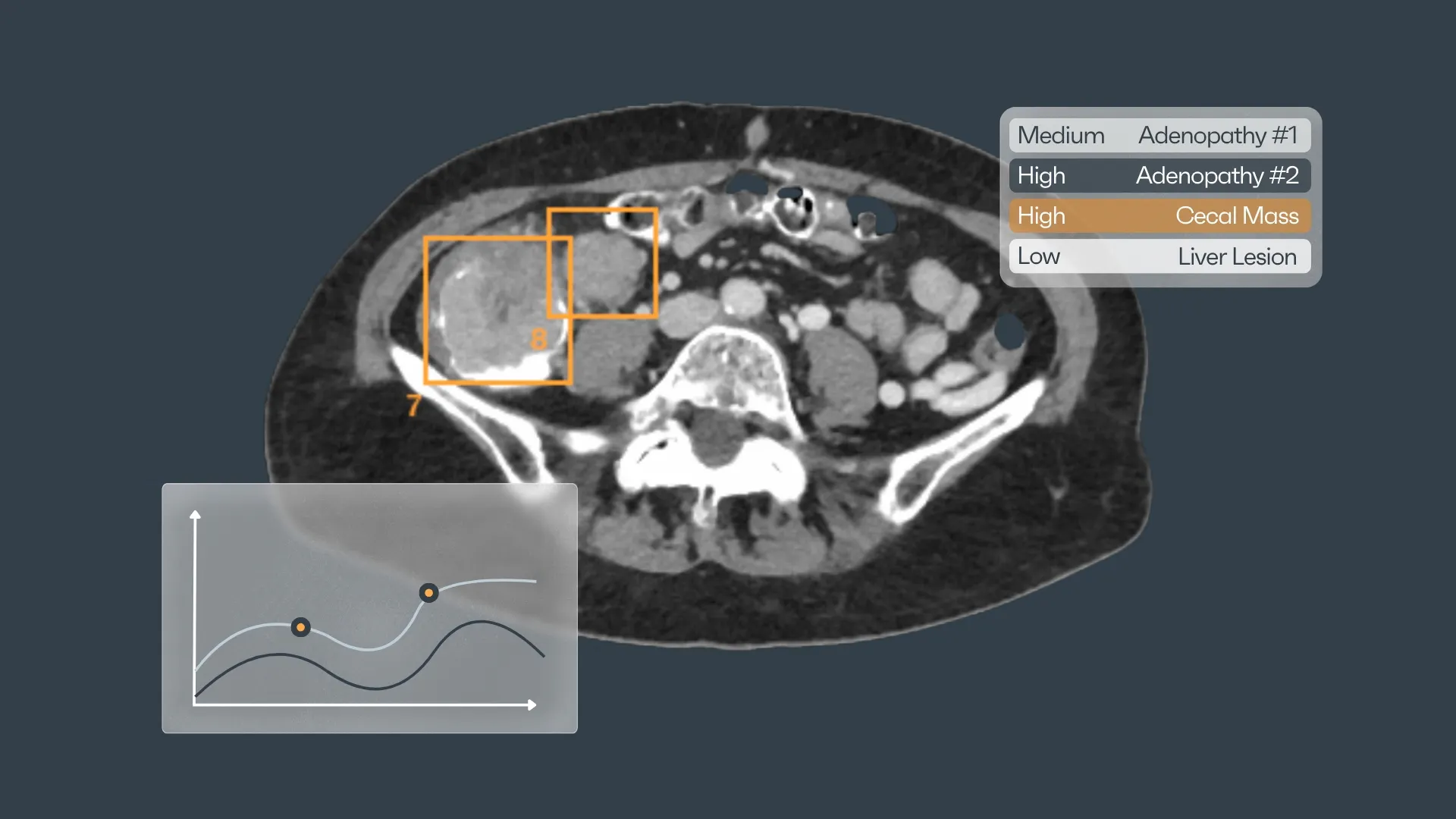
This is where we see huge potential for AI to help us—not just as detectors, but as systems that can think longitudinally. Tools that remember what we might forget across dozens of cases. That can automatically highlight relevant changes from prior studies, which understand the narrative arc of a patient's disease journey. The real difficulty lies in the meaning that threads them together.
Radiologists don’t see pixels; they see physiology in motion
Each voxel of attenuation represents competing biological forces: perfusion, necrosis, inflammation, and fibrosis. What the algorithm labels as “lesion” might, in truth, be the aftermath of therapy, the ghost of a tumor that once was. To discern the living from the dead tissue, we interpret change across time, modalities, and context. That temporal reasoning, the ability to understand cause and consequence, is what separates pattern recognition from true clinical understanding. For AI to assist in oncology, it must learn not only to see, but to remember and to compare.
This temporal and spatial integration challenges even human cognition. Consider the patient who undergoes chemotherapy, immunotherapy, and radiation in rapid succession. The scans evolve: what once enhanced avidly now fades; what once was invisible becomes conspicuous. The radiologist must interpret this visual symphony while recalling prior studies, treatment regimens, and clinical symptoms. For an algorithm, this means bridging structured data with unstructured narrative, correlating images with evolving therapies, and adjusting interpretations dynamically. So, when we try to build AI for oncology imaging, we are not asking for a faster detector; we are asking for a companion that can think longitudinally. We are asking it to hold uncertainty, to weigh the improbable, to understand that in cancer, paradoxes are common and certainty is dangerous. The success of such intelligence will not be measured merely by accuracy metrics, but by its ability to preserve the radiologist’s capacity for clinical imagination, to illuminate patterns without dulling the human intuition that has always been at the heart of medicine.
What radiologists actually need is a shift in architecture. Current AI solutions are like a series of specialists, each trained on a narrow task: one for lung nodules, another for bone lesions, etc. Each specialist performs well in isolation, but none understands the patient as a whole. The next generation of oncology imaging AI must resemble a generalist: a model that learns across modalities and organs, synthesizing disparate inputs into a coherent picture.
So what does the ideal oncology AI look like? Let us share what would make the biggest difference in my work:
- Systems that see patients holistically. Tools that can analyze multiple body regions together and understand systemic relationships. That recognizes when findings across different organs might be telling a connected story.
- Intelligence that understands time. Platforms that automatically contextualize findings against prior studies. That helps distinguish real progression from treatment effect. That knows when follow-up imaging is due based on protocols.
- Outputs that speak clinical language. Not just confidence scores and bounding boxes, but narratives and structured reports that make sense to the oncology team. "Three new liver lesions compared to two months ago, suggesting progression despite therapy"—that's the kind of synthesis that drives clinical decisions.
- Transparency about limitations. Systems that tell us when findings are in gray zones requiring human judgment. That doesn't oversell certainty on ambiguous cases. That knows some questions need biopsy, not just better imaging.
But getting from here to there requires real collaboration between technologists and clinicians. Solutions need to understand not just what we do, but how we think. How we approach complex cases. What questions keep us up at night? And the clinicians should engage with this technology thoughtfully—articulating what they actually need, testing what works in real-world practice, and providing feedback that helps developers build better tools.
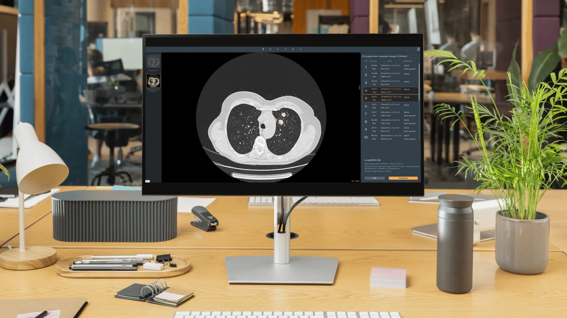
This holistic future is both ambitious and necessary, but as cancer does not confine itself to a single organ, neither should our tools for detecting and managing it. A unified oncology imaging AI, one that understands systemic relationships, that tells coherent stories and recognizes patterns across modalities, this should be the promise of the companies in the industry, to make diagnosis more timely, treatment more precise, and follow‑up more reliable. This is not an abstract dream but a direction already charted, and the work has just begun.
If this perspective resonates, if you share the curiosity for an intelligence that can truly understand the body’s story, we invite you to connect, exchange ideas, and take part in shaping the next chapter of AI imaging.


Discover how Rayscape works
integrate Rayscape AI into
your hospital's infrastructure
and improve both your
daily workflow and patients' experience.
