

Abdominal CT AI and the Challenge of Early Cancer Detection
An abdominal CT is not a single dataset; it is a layered density problem. Each organ behaves according to its own physics. The liver demonstrates gradual enhancement and coarse parenchymal echotexture. The spleen shows its characteristic mottled enhancement. The pancreas alternates between lobulated glandular tissue and peripancreatic fat. Kidneys cycle through corticomedullary, nephrographic, and excretory patterns. Meanwhile, the bowel loops introduce motion, fluid, gas, partial volume effects, and unpredictable contrast patterns. All of these elements coexist within a limited anatomical volume, forcing radiologists to interpret dozens of competing signals at once.
Cancer grows inside this density chaos. Early lesions often produce only minor deviations from expected attenuation — sometimes a change of three to five Hounsfield Units compared to the surrounding tissue. Small hepatic metastases may be nearly isodense on the portal venous phase and visible only in a fleeting arterial window. Subtle renal masses can hide until the nephrographic phase equalizes their attenuation. Pancreatic adenocarcinoma may not create a discrete mass at all, but instead a barely perceptible hypoattenuating focus or secondary signs such as abrupt ductal cutoff.
Where complexity begins: anatomy and density that defy simple rules
This is the reason rule-based systems have historically failed in abdominal CT. There is no consistent pattern that applies across organs. Texture, enhancement kinetics, and lesion morphology shift dramatically from region to region. A “solid lesion” is not a universal category; it is a context-dependent phenomenon. A hypervascular nodule in the liver behaves differently from a hypervascular cortical mass in the kidney. Hypodense pancreatic foci follow entirely different enhancement rules than hypodense splenic lesions.
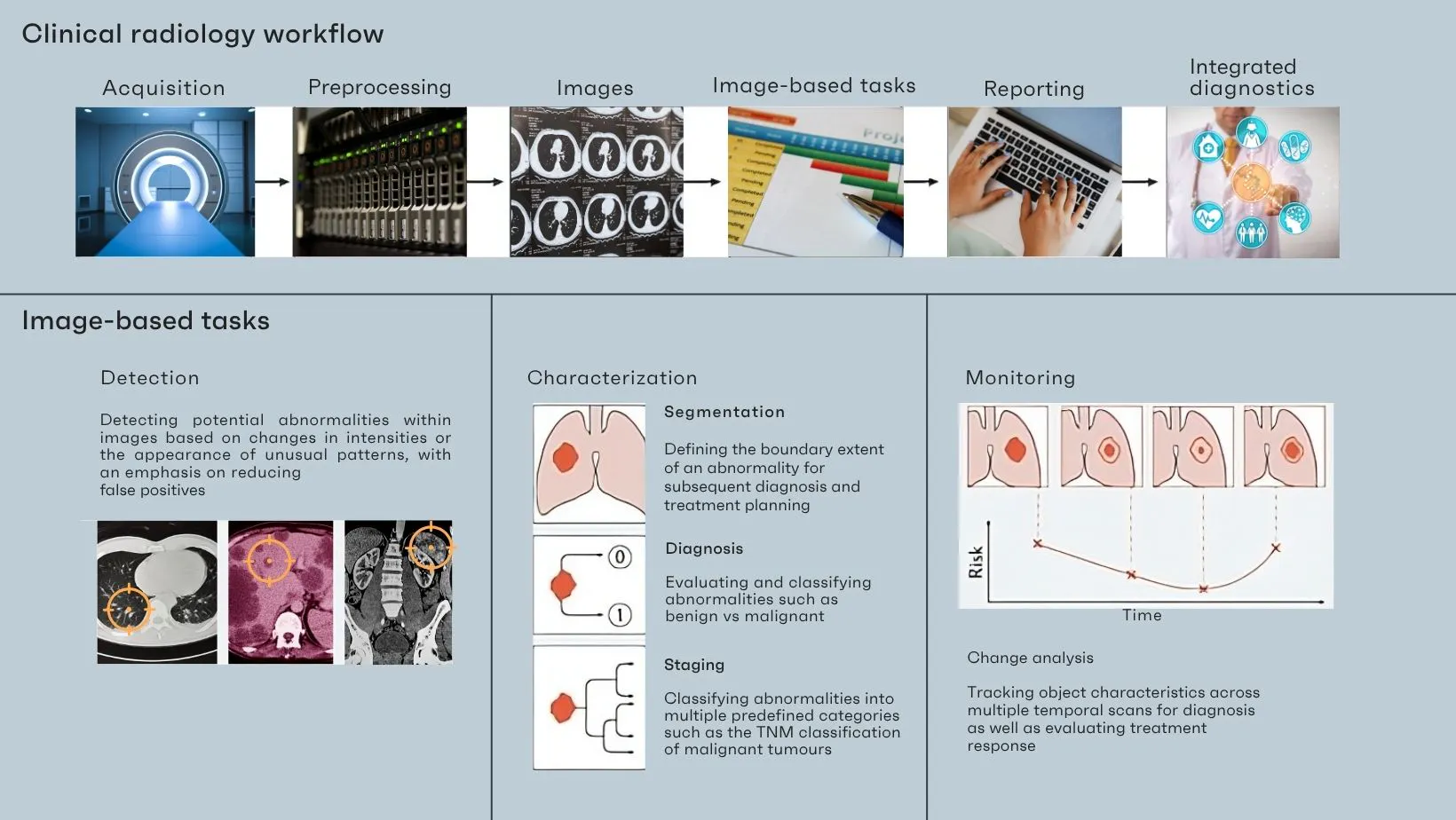
The fat planes that should theoretically separate structures often mislead. Inflammation, edema, desmoplastic reaction, and prior surgery distort expected boundaries. Density clusters that appear pathologic in one organ are normal variants in another. Partial volume effects mimic pathology, especially at organ interfaces.
Even deep-learning models trained successfully on thoracic CT struggle when confronted with this heterogeneity. Thoracic datasets have clearer density stratification and less motion; abdominal datasets contain overlapping textures, vascular pulsation, respiratory artifacts, and highly variable enhancement. The network must learn not simply to detect lesions but to understand how each organ behaves across multiple phases, a task that requires far more data, far more nuance, and far richer annotation.
The abdomen resists simplification because its signals are entangled. Anatomy is complex, density is relativistic, and pathology is subtle. This is the maze radiologists navigate every day — and the environment any AI model must learn to interpret before it can meaningfully assist in cancer detection.
Overlapping pathologies and early lesions that vanish in plain sight
If abdominal anatomy is a geometric puzzle, its pathology is a master of disguise. Many malignancies arise not as discrete masses but as subtle distortions of normal physiology, hiding within enhancement patterns that radiologists already know are highly variable from patient to patient.
Pancreatic adenocarcinoma may be the clearest example. Early tumors frequently produce no definable mass; they present instead as a vague hypoattenuating region that mirrors the gland’s natural lobulated texture. The background heterogeneity of the pancreas, influenced by age, fat infiltration, and contrast timing, becomes the perfect camouflage. Sometimes the only real clues are secondary phenomena: an abrupt pancreatic duct cutoff, upstream ductal dilation, or loss of the normal “marbled” enhancement. These are findings that require an experienced eye and are easy to overlook during a busy read.
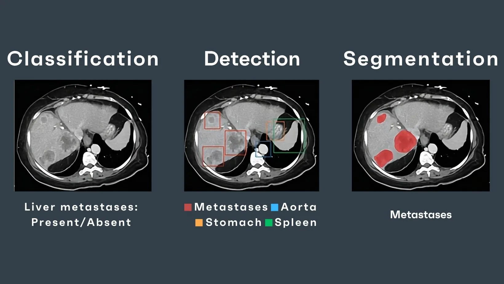
Hepatic metastases play their own game of mimicry. Depending on vascularity and contrast phase, the same lesion can imitate a benign cyst, a flash-filling hemangioma, or nothing at all. Hypervascular metastases may be conspicuous in the arterial phase but nearly invisible in the portal venous phase; hypovascular metastases behave in the opposite direction. Lesions that are technically present on a given phase may not reach the conspicuity threshold needed for confident detection.
Radiologists encounter these traps daily, which is why abdominal CT second opinions are so common and why early malignant lesions across the thoracoabdominopelvic axis are among the most commonly missed findings in retrospective audits. The challenge is not a lack of skill but the inherent subtlety of early disease. Without multi-phase acquisition: arterial, portal venous, delayed, even experienced readers revisit scans with the uneasy sense that a lesion may have slipped beneath the threshold of detection.
AI inherits this environment without any shortcuts. A model can only recognize what it has seen and internalized, and early-stage lesions sit exactly at the boundary between meaningful signal and background noise. The statistical distinction between a tiny implant and a random fat striation is razor-thin. In regions where radiologists already rely on context, comparison, experience, and multimodal reasoning, an algorithm must attempt to extract patterns from very weak gradients.
This is where sensitivity dips, especially for small, isodense, or phase-dependent lesions. And this is also where false positives emerge, as benign structures, enhancing spleen nodules, perfusion variations, focal fatty sparing, and hyperdense bowel content mimic malignancy too well for a model that lacks full clinical context.
The difficulty lies not in dramatic tumors but in the ones that seem to vanish inside the very physiology they disrupt, and AI must learn to hear those whispers before it can meaningfully assist the radiologist.
How AI can help, not by replacing vision, but by expanding it
The promise of AI in abdominal TAP oncology is not a cinematic transformation but an incremental one. Multi-region and multi-organ scans can be too complex, the pathological spectrum too broad, and the density interactions too subtle for any algorithmic leap to “solve” cancer detection outright. Progress comes from refinement: amplifying weak signals, stabilizing variability, and giving radiologists additional layers of perceptual and analytical support.
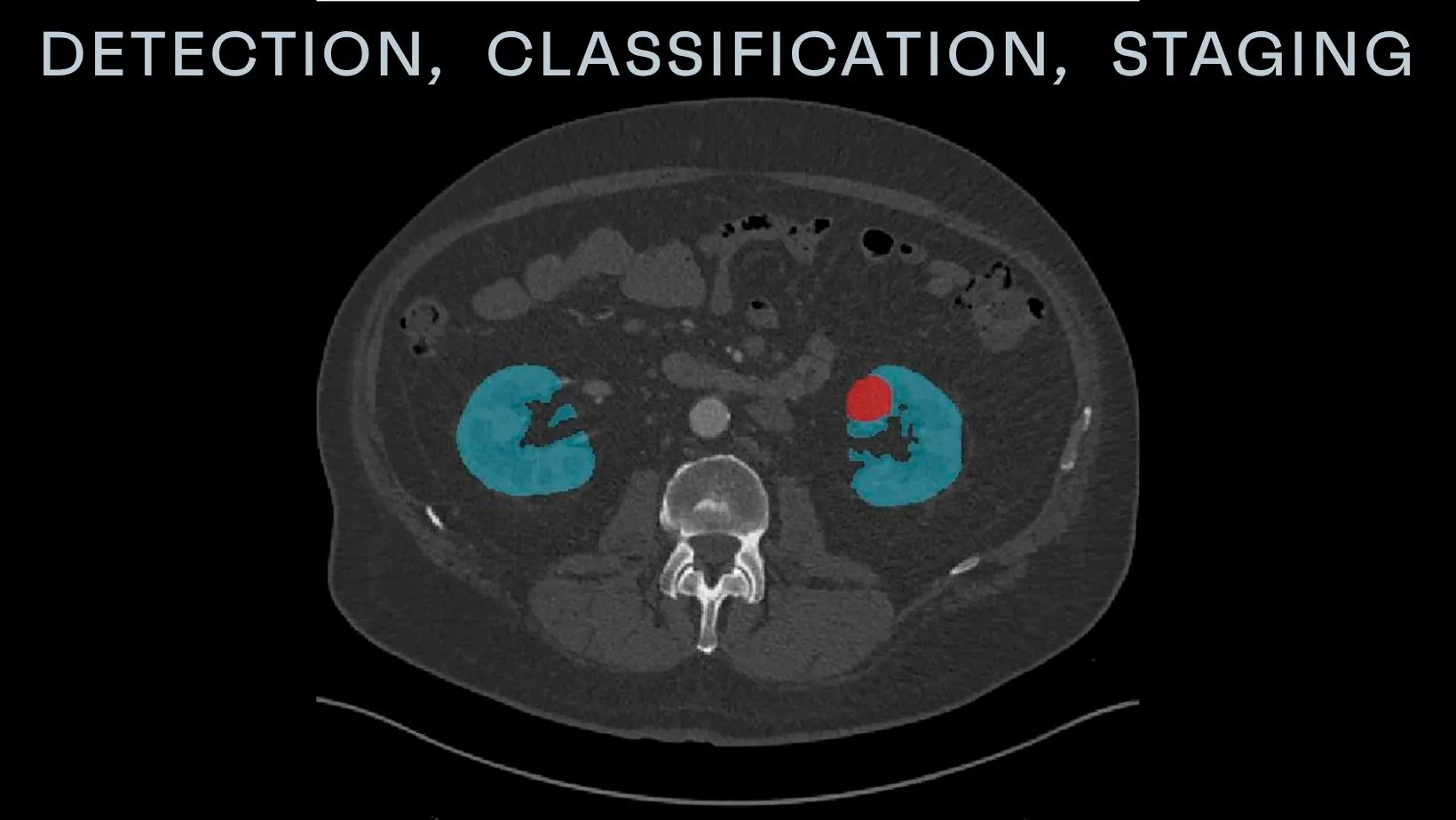
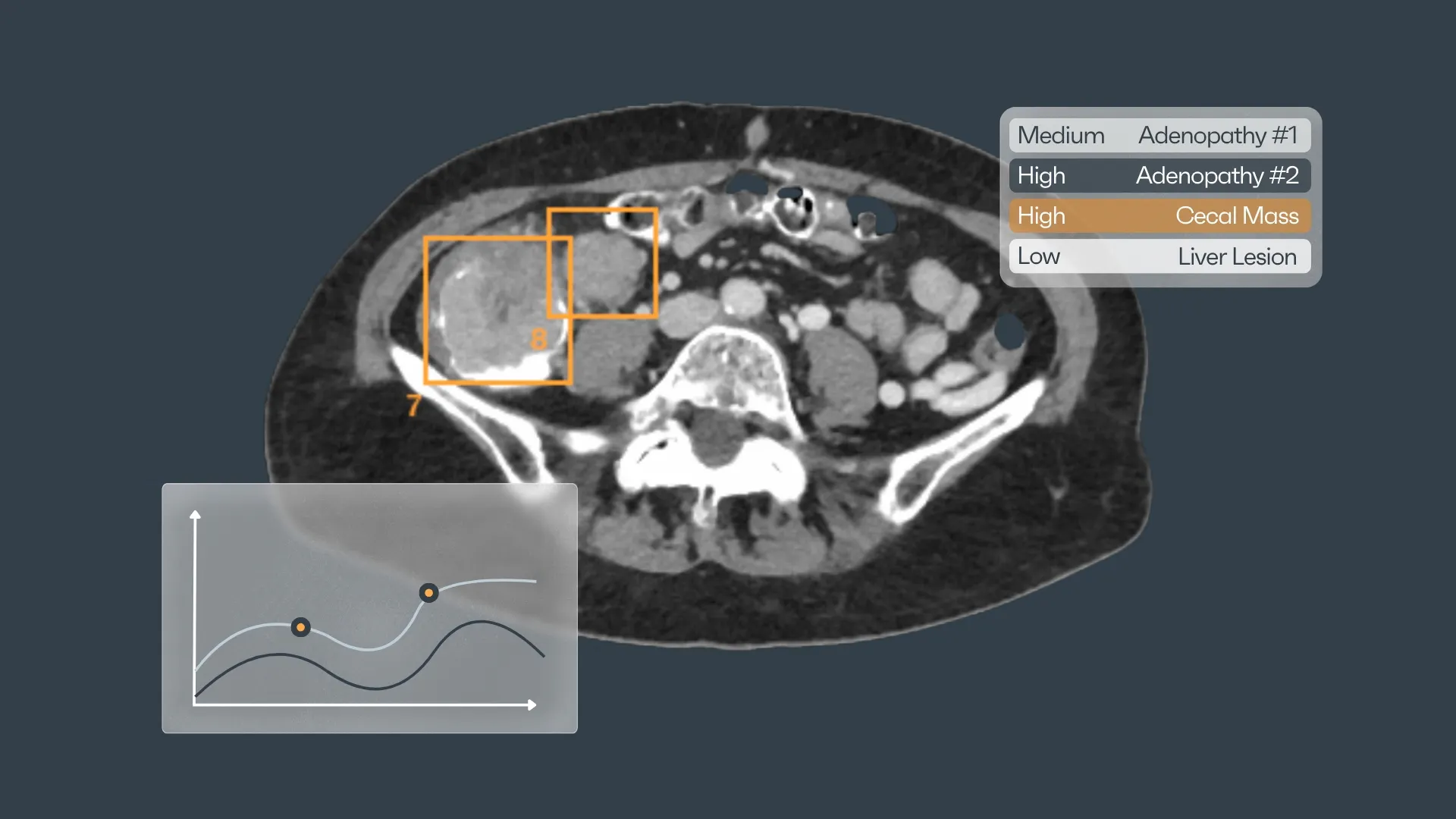
Modern abdominal CT AI does not interpret a slice in isolation. It processes the dataset as a volumetric continuum, evaluating tissue relationships voxel by voxel. Instead of relying solely on macroscopic morphology, it quantifies micro-patterns: textural entropy, attenuation gradients, vascular signatures, and geometric deviations that fall below the human perceptual threshold. These statistical fingerprints often capture the earliest signs of malignant transformation, shifts that remain invisible to the naked eye because they do not yet form a mass or cause architectural distortion.
Where a radiologist might see a slightly irregular contour of the liver capsule, the algorithm can measure whether that contour deviates significantly from expected anatomical curvature. Where the eye sees a faint hypoattenuating patch in the pancreas, the model evaluates whether the texture, enhancement kinetics, and local ductal configuration match thousands of prior examples of early adenocarcinoma. The human brain compares images; AI computes distributions.
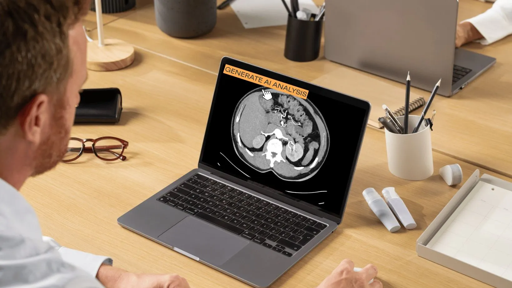
The value of this computational perspective is not to override radiologic judgment but to complement it. AI can flag inconsistencies that warrant a second look, subtle lesions that might otherwise be dismissed as noise or artifact. It can highlight alternative differentials in regions where benign and malignant entities overlap in appearance. In follow-up imaging, AI quantifies lesion growth with millimetric precision, reducing cognitive bias and helping clinicians distinguish true progression from the illusion of progression caused by slice positioning or partial volume.
This kind of assistance matters profoundly in abdominal and pelvic imaging. A flagged 8-mm lesion that prompts re-evaluation can lead to earlier diagnosis. A reminder to reconsider a pancreatic contour change may alter patient management. A consistent volumetric tracking system can transform ambiguous follow-up intervals into actionable clinical insight.
In a domain defined by complexity, the role of AI is to widen the radiologist’s perceptual bandwidth. It surfaces weak signals, stabilizes interpretation across phases, and introduces a layer of mathematical consistency that the human visual system alone cannot sustain.
The abdominal maze becomes more navigable when human and artificial intelligence work in parallel, each compensating for the other’s blind spots.
From regional expertise to whole-body insight, a natural progression
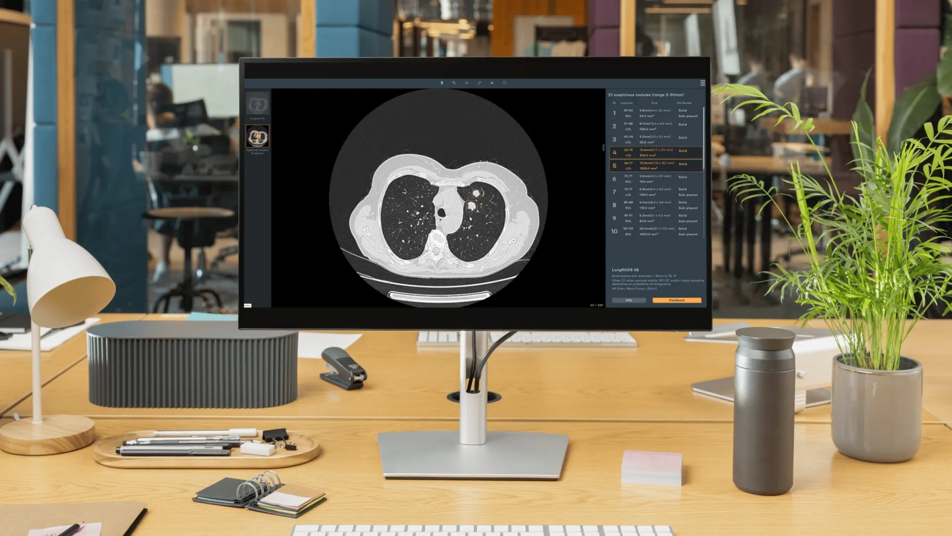
Rayscape’s foundation in thoracic imaging has given us years of practical exposure to how AI behaves in real clinical environments. Chest CT was a laboratory for understanding the mechanics of radiologic complexity itself. In the lungs, we learned how models cope with motion, variable aeration, overlapping patterns of structures, and the challenge of distinguishing true pathology from artifacts or benign variants. We studied how subtle textural changes can precede overt mass formation, how contrast dynamics influence conspicuity, and how AI performance shifts across scanners, protocols, and patient populations.
These experiences taught us something deeper: AI becomes clinically meaningful not through isolated detections, but through consistent interpretation across thousands of heterogeneous cases. Radiologists respond to stability, reproducibility, and transparency. Rayscape LungCT gave us the data to refine those principles. It showed us how AI must operate in regions where normal variation is broad, early lesions are faint, and disease rarely conforms to neat visual categories.
This foundation naturally points toward extension. Oncologic imaging does not begin and end at the diaphragm; it follows patterns of spread, vascular pathways, lymphatic chains, and organ interfaces. The analytic strategies we refined in thoracic work: volumetric modeling, sensitivity to micro-attenuation changes, cross-phase reasoning, and pattern recognition, are the same strategies required to interpret malignancy in the liver, pancreas, retroperitoneum, peritoneal surfaces, or other organs.
Expanding into these regions is not a conceptual leap so much as an extension of what radiologists already do: synthesizing information across organ systems, integrating subtle clues from multiple compartments, and tracking disease as a continuum rather than a set of isolated findings. Rayscape’s ongoing exploration of oncology imaging AI follows that same logic. With each dataset, annotation, and clinical partnership, we accumulate the domain knowledge necessary to approach these anatomically dense and diagnostically subtle regions with the same scientific rigor that defined our work in the chest.
For teams exploring how advanced imaging intelligence can support their radiologists, streamline interpretation, or enhance oncologic workflows, Rayscape is already working in these spaces. If you’re interested in discussing our current AI solutions or learning more about our ongoing work in thoracic and oncologic imaging, feel free to reach out. We’re always open to thoughtful conversations with clinicians and institutions shaping the future of radiology.


Discover how Rayscape works
integrate Rayscape AI into
your hospital's infrastructure
and improve both your
daily workflow and patients' experience.
